2W97

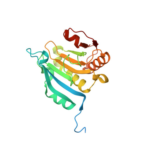
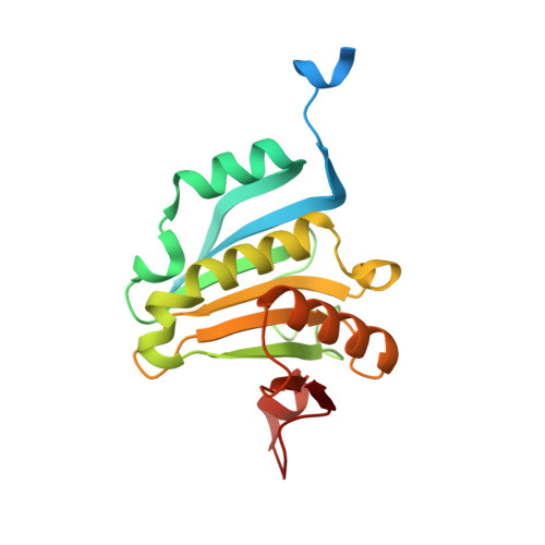
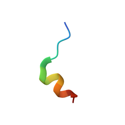
Crystal Structure of eIF4E Bound to Glycerol and eIF4G1 peptide


Experimental Data Snapshot

  • Method: X-RAY DIFFRACTION
  • Resolution: 2.29 Å
  • R-Value Free: 0.239 
  • R-Value Work: 0.197 
  • R-Value Observed: 0.199 

Starting Model: experimental
View more details

wwPDB Validation   3D Report Full Report


Ligand Structure Quality Assessment 


This is version 1.4 of the entry. See complete history


Literature

Crystallization of eIF4E complexed with eIF4GI peptide and glycerol reveals distinct structural differences around the cap-binding site.

Brown, C.J.Verma, C.S.Walkinshaw, M.D.Lane, D.P.

(2009) Cell Cycle 8: 1905-1911

  • DOI: https://doi.org/10.4161/cc.8.12.8742
  • Primary Citation of Related Structures:  
    2W97

  • PubMed Abstract: 

    An X-ray crystal structure of the eIF4E peptide complex is described in which two such complexes are located in the asymmetric unit. One of these complexes has m(7)GTP bound in a conformation which has been observed in several eIF4E crystal structures, whilst the other complex is free of m(7)GTP and contains a unique glycerol. The two complexes show significant structural differences between each other in the cap-binding site. The glycerol bound structure shows a reorientation of the W102 side chain out of the cap-binding site, disordering of the W56 containing loop and rotation of the carboxyl side-chain of E103. This is accompanied by movement of the M101 side chain into a position where W56 in the m(7)GTP bound complex would otherwise occupy. Rotation of the W102 sidechain also displaces a structured water molecule to a new site. This novel conformation of eIF4E with glycerol bound is hypothesized to be an intermediate state between the apo and m(7)GTP bound forms of eIF4E. These insights should prove useful in the design of inhibitors of eIF4E for cancer therapy.


  • Organizational Affiliation

    Institute of Molecular and Cell Biology (A-STAR), Proteos, Singapore.


Macromolecules
Find similar proteins by:  (by identity cutoff)  |  3D Structure
Entity ID: 1
MoleculeChains Sequence LengthOrganismDetailsImage
EUKARYOTIC TRANSLATION INITIATION FACTOR 4E217Homo sapiensMutation(s): 0 
UniProt & NIH Common Fund Data Resources
Find proteins for P06730 (Homo sapiens)
Explore P06730 
Go to UniProtKB:  P06730
PHAROS:  P06730
GTEx:  ENSG00000151247 
Entity Groups  
Sequence Clusters30% Identity50% Identity70% Identity90% Identity95% Identity100% Identity
UniProt GroupP06730
Sequence Annotations
Expand
  • Reference Sequence
Find similar proteins by:  (by identity cutoff)  |  3D Structure
Entity ID: 2
MoleculeChains Sequence LengthOrganismDetailsImage
EUKARYOTIC TRANSLATION INITIATION FACTOR 4E217Homo sapiensMutation(s): 0 
UniProt & NIH Common Fund Data Resources
Find proteins for P06730 (Homo sapiens)
Explore P06730 
Go to UniProtKB:  P06730
PHAROS:  P06730
GTEx:  ENSG00000151247 
Entity Groups  
Sequence Clusters30% Identity50% Identity70% Identity90% Identity95% Identity100% Identity
UniProt GroupP06730
Sequence Annotations
Expand
  • Reference Sequence

Find similar proteins by:  Sequence   |   3D Structure  

Entity ID: 3
MoleculeChains Sequence LengthOrganismDetailsImage
EUKARYOTIC TRANSLATION INITIATION FACTOR 4 GAMMA 1C [auth E],
D [auth F]
14Homo sapiensMutation(s): 0 
UniProt & NIH Common Fund Data Resources
Find proteins for Q04637 (Homo sapiens)
Explore Q04637 
Go to UniProtKB:  Q04637
PHAROS:  Q04637
GTEx:  ENSG00000114867 
Entity Groups  
Sequence Clusters30% Identity50% Identity70% Identity90% Identity95% Identity100% Identity
UniProt GroupQ04637
Sequence Annotations
Expand
  • Reference Sequence
Small Molecules
Ligands 4 Unique
IDChains Name / Formula / InChI Key2D Diagram3D Interactions
MGO
Query on MGO

Download Ideal Coordinates CCD File 
E [auth A][[(2R,3S,4R,5R)-5-(6-AMINO-3-METHYL-4-OXO-5H-IMIDAZO[4,5-C]PYRIDIN-1-YL)-3,4-DIHYDROXY-OXOLAN-2-YL]METHOXY-HYDROXY-PHOSPHORYL] PHOSPHONO HYDROGEN PHOSPHATE
C12 H20 N4 O14 P3
LPHBJZOLBGBNJF-XYHAGOFUSA-O
PGE
Query on PGE

Download Ideal Coordinates CCD File 
F [auth A]TRIETHYLENE GLYCOL
C6 H14 O4
ZIBGPFATKBEMQZ-UHFFFAOYSA-N
SO4
Query on SO4

Download Ideal Coordinates CCD File 
G [auth A],
H [auth A],
J [auth B],
K [auth B]
SULFATE ION
O4 S
QAOWNCQODCNURD-UHFFFAOYSA-L
GOL
Query on GOL

Download Ideal Coordinates CCD File 
I [auth A],
L [auth B]
GLYCEROL
C3 H8 O3
PEDCQBHIVMGVHV-UHFFFAOYSA-N
Binding Affinity Annotations 
IDSourceBinding Affinity
GOL PDBBind:  2W97 Kd: 1.10e+8 (nM) from 1 assay(s)
Experimental Data & Validation

Experimental Data

  • Method: X-RAY DIFFRACTION
  • Resolution: 2.29 Å
  • R-Value Free: 0.239 
  • R-Value Work: 0.197 
  • R-Value Observed: 0.199 
  • Space Group: P 1 21 1
Unit Cell:
Length ( Å )Angle ( ˚ )
a = 84.106α = 90
b = 38.201β = 101.51
c = 93.687γ = 90
Software Package:
Software NamePurpose
REFMACrefinement
HKL-2000data reduction
HKL-2000data scaling
PHASERphasing

Structure Validation

View Full Validation Report


Ligand Structure Quality Assessment 


Entry History 

Deposition Data

Revision History  (Full details and data files)

  • Version 1.0: 2010-03-31
    Type: Initial release
  • Version 1.1: 2011-05-08
    Changes: Version format compliance
  • Version 1.2: 2011-07-13
    Changes: Version format compliance
  • Version 1.3: 2019-10-09
    Changes: Data collection, Database references, Other
  • Version 1.4: 2023-12-13
    Changes: Data collection, Database references, Derived calculations, Refinement description