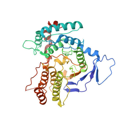
2WVM

H309A mutant of Mannosyl-3-phosphoglycerate synthase from Thermus thermophilus HB27 in complex with GDP-alpha-D-Mannose and Mg(II)


Experimental Data Snapshot

  • Method: X-RAY DIFFRACTION
  • Resolution: 2.98 Å
  • R-Value Free: 0.232 
  • R-Value Work: 0.174 
  • R-Value Observed: 0.176 

Starting Model: experimental
View more details

wwPDB Validation   3D Report Full Report


Ligand Structure Quality Assessment 


This is version 1.3 of the entry. See complete history


Literature

Structural Analysis of Thermus Thermophilus Hb27 Mannosyl-3-Phosphoglycerate Synthase Provides Evidence for a Second Catalytic Metal Ion and New Insight Into the Retaining Mechanism of Glycosyltransferases.

Goncalves, S.Borges, N.Esteves, A.M.Victor, B.Soares, C.M.Santos, H.Matias, P.M.

(2010) J Biol Chem 285: 17857

  • DOI: https://doi.org/10.1074/jbc.M109.095976
  • Primary Citation of Related Structures:  
    2WVK, 2WVL, 2WVM

  • PubMed Abstract: 

    Mannosyl-3-phosphoglycerate synthase is a glycosyltransferase involved in the two-step synthetic pathway of mannosylglycerate, a compatible solute that accumulates in response to salt and/or heat stresses in many microorganisms thriving in hot environments. The three-dimensional structure of mannosyl-3-phosphoglycerate synthase from Thermus thermophilus HB27 in its binary complex form, with GDP-alpha-D-mannose and Mg(2+), shows a second metal binding site, about 6 A away from the mannose moiety. Kinetic and mutagenesis studies have shown that this metal site plays a role in catalysis. Additionally, Asp(167) in the DXD motif is found within van der Waals contact distance of the C1' atom in the mannopyranose ring, suggesting its action as a catalytic nucleophile, either in the formation of a glycosyl-enzyme intermediate according to the double-displacement S(N)2 reaction mechanism or in the stabilization of the oxocarbenium ion-like intermediate according to the D(N)*A(Nss) (S(N)i-like) reaction mechanism. We propose that either mechanism may occur in retaining glycosyltransferases with a GT-A fold, and, based on the gathered structural information, we identified an extended structural signature toward a common scaffold between the inverting and retaining glycosyltransferases.


  • Organizational Affiliation

    Instituto de Tecnologia Química e Biológica, Universidade Nova de Lisboa, Apartado 127, 2781-901 Oeiras, Portugal.


Macromolecules
Find similar proteins by:  (by identity cutoff)  |  3D Structure
Entity ID: 1
MoleculeChains Sequence LengthOrganismDetailsImage
MANNOSYL-3-PHOSPHOGLYCERATE SYNTHASE
A, B
391Thermus thermophilus HB27Mutation(s): 1 
EC: 2.4.1.217 (PDB Primary Data), 2.4.1 (UniProt)
UniProt
Find proteins for Q72K30 (Thermus thermophilus (strain ATCC BAA-163 / DSM 7039 / HB27))
Explore Q72K30 
Go to UniProtKB:  Q72K30
Entity Groups  
Sequence Clusters30% Identity50% Identity70% Identity90% Identity95% Identity100% Identity
UniProt GroupQ72K30
Sequence Annotations
Expand
  • Reference Sequence
Small Molecules
Ligands 3 Unique
IDChains Name / Formula / InChI Key2D Diagram3D Interactions
GDD
Query on GDD

Download Ideal Coordinates CCD File 
C [auth A],
F [auth B]
GUANOSINE-5'-DIPHOSPHATE-ALPHA-D-MANNOSE
C16 H25 N5 O16 P2
MVMSCBBUIHUTGJ-GDJBGNAASA-N
ZN
Query on ZN

Download Ideal Coordinates CCD File 
E [auth A],
H [auth B],
I [auth B]
ZINC ION
Zn
PTFCDOFLOPIGGS-UHFFFAOYSA-N
MG
Query on MG

Download Ideal Coordinates CCD File 
D [auth A],
G [auth B]
MAGNESIUM ION
Mg
JLVVSXFLKOJNIY-UHFFFAOYSA-N
Experimental Data & Validation

Experimental Data

  • Method: X-RAY DIFFRACTION
  • Resolution: 2.98 Å
  • R-Value Free: 0.232 
  • R-Value Work: 0.174 
  • R-Value Observed: 0.176 
  • Space Group: P 41 21 2
Unit Cell:
Length ( Å )Angle ( ˚ )
a = 112.766α = 90
b = 112.766β = 90
c = 199.69γ = 90
Software Package:
Software NamePurpose
REFMACrefinement
XDSdata reduction
XDSdata scaling
PHASERphasing

Structure Validation

View Full Validation Report


Ligand Structure Quality Assessment 


Entry History 

Deposition Data

Revision History  (Full details and data files)

  • Version 1.0: 2010-03-31
    Type: Initial release
  • Version 1.1: 2011-07-13
    Changes: Advisory, Version format compliance
  • Version 1.2: 2014-02-19
    Changes: Database references, Source and taxonomy
  • Version 1.3: 2023-12-20
    Changes: Data collection, Database references, Derived calculations, Other, Refinement description