2XZ4 | pdb_00002xz4

Crystal structure of the LFZ ectodomain of the peptidoglycan recognition protein LF


Experimental Data Snapshot

  • Method: X-RAY DIFFRACTION
  • Resolution: 1.72 Å
  • R-Value Free: 
    0.201 (Depositor), 0.190 (DCC) 
  • R-Value Work: 
    0.164 (Depositor), 0.160 (DCC) 
  • R-Value Observed: 
    0.166 (Depositor) 

Starting Model: experimental
View more details

wwPDB Validation   3D Report Full Report


This is version 1.4 of the entry. See complete history


Literature

The Drosophila Peptidoglycan-Recognition Protein Lf Interacts with Peptidoglycan-Recognition Protein Lc to Downregulate the Imd Pathway.

Basbous, N.Coste, F.Leone, P.Vincentelli, R.Royet, J.Kellenberger, C.Roussel, A.

(2011) EMBO Rep 12: 327

  • DOI: https://doi.org/10.1038/embor.2011.19
  • Primary Citation of Related Structures:  
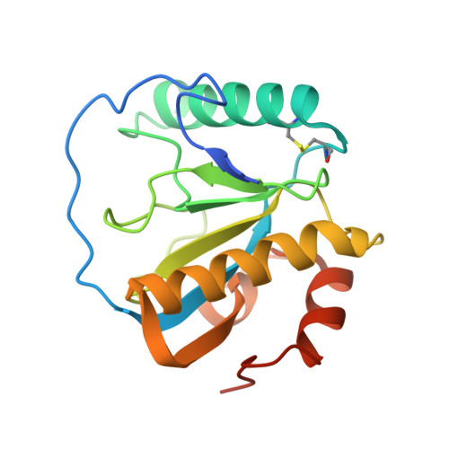
    2XZ4, 2XZ8

  • PubMed Abstract: 

    The peptidoglycan (PGN)-recognition protein LF (PGRP-LF) is a specific negative regulator of the immune deficiency (Imd) pathway in Drosophila. We determine the crystal structure of the two PGRP domains constituting the ectodomain of PGRP-LF at 1.72 and 1.94 Å resolution. The structures show that the LFz and LFw domains do not have a PGN-docking groove that is found in other PGRP domains, and they cannot directly interact with PGN, as confirmed by biochemical-binding assays. By using surface plasmon resonance analysis, we show that the PGRP-LF ectodomain interacts with the PGRP-LCx ectodomain in the absence and presence of tracheal cytotoxin. Our results suggest a mechanism for downregulation of the Imd pathway on the basis of the competition between PRGP-LCa and PGRP-LF to bind to PGRP-LCx.


  • Organizational Affiliation

    Centre de Biophysique Moléculaire, UPR 4301 CNRS, Rue Charles Sadron, Orléans 2 45071, France.


Macromolecules
Find similar proteins by:  (by identity cutoff)  |  3D Structure
Entity ID: 1
MoleculeChains Sequence LengthOrganismDetailsImage
PEPTIDOGLYCAN-RECOGNITION PROTEIN LF
A, B
180Drosophila melanogasterMutation(s): 0 
UniProt
Find proteins for Q8SXQ7 (Drosophila melanogaster)
Explore Q8SXQ7 
Go to UniProtKB:  Q8SXQ7
Entity Groups  
Sequence Clusters30% Identity50% Identity70% Identity90% Identity95% Identity100% Identity
UniProt GroupQ8SXQ7
Sequence Annotations
Expand
  • Reference Sequence
Small Molecules
Ligands 3 Unique
IDChains Name / Formula / InChI Key2D Diagram3D Interactions
1PG
Query on 1PG

Download Ideal Coordinates CCD File 
F [auth A]2-(2-{2-[2-(2-METHOXY-ETHOXY)-ETHOXY]-ETHOXY}-ETHOXY)-ETHANOL
C11 H24 O6
SLNYBUIEAMRFSZ-UHFFFAOYSA-N
CU
Query on CU

Download Ideal Coordinates CCD File 
C [auth A]COPPER (II) ION
Cu
JPVYNHNXODAKFH-UHFFFAOYSA-N
EDO
Query on EDO

Download Ideal Coordinates CCD File 
D [auth A],
E [auth A],
G [auth B],
H [auth B],
I [auth B]
1,2-ETHANEDIOL
C2 H6 O2
LYCAIKOWRPUZTN-UHFFFAOYSA-N
Experimental Data & Validation

Experimental Data

  • Method: X-RAY DIFFRACTION
  • Resolution: 1.72 Å
  • R-Value Free:  0.201 (Depositor), 0.190 (DCC) 
  • R-Value Work:  0.164 (Depositor), 0.160 (DCC) 
  • R-Value Observed: 0.166 (Depositor) 
Space Group: P 21 21 2
Unit Cell:
Length ( Å )Angle ( ˚ )
a = 74.802α = 90
b = 113.443β = 90
c = 37.532γ = 90
Software Package:
Software NamePurpose
PHENIXrefinement
iMOSFLMdata reduction
SCALAdata scaling
PHENIXphasing

Structure Validation

View Full Validation Report


Entry History 

Deposition Data

Revision History  (Full details and data files)

  • Version 1.0: 2011-04-13
    Type: Initial release
  • Version 1.1: 2011-05-08
    Changes: Version format compliance
  • Version 1.2: 2011-07-13
    Changes: Version format compliance
  • Version 1.3: 2023-12-20
    Changes: Data collection, Database references, Derived calculations, Other, Refinement description
  • Version 1.4: 2024-11-13
    Changes: Structure summary