2YR4

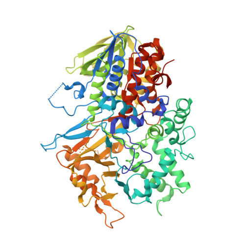
Crystal structure of L-phenylalanine oxiase from Psuedomonas sp. P-501


Experimental Data Snapshot

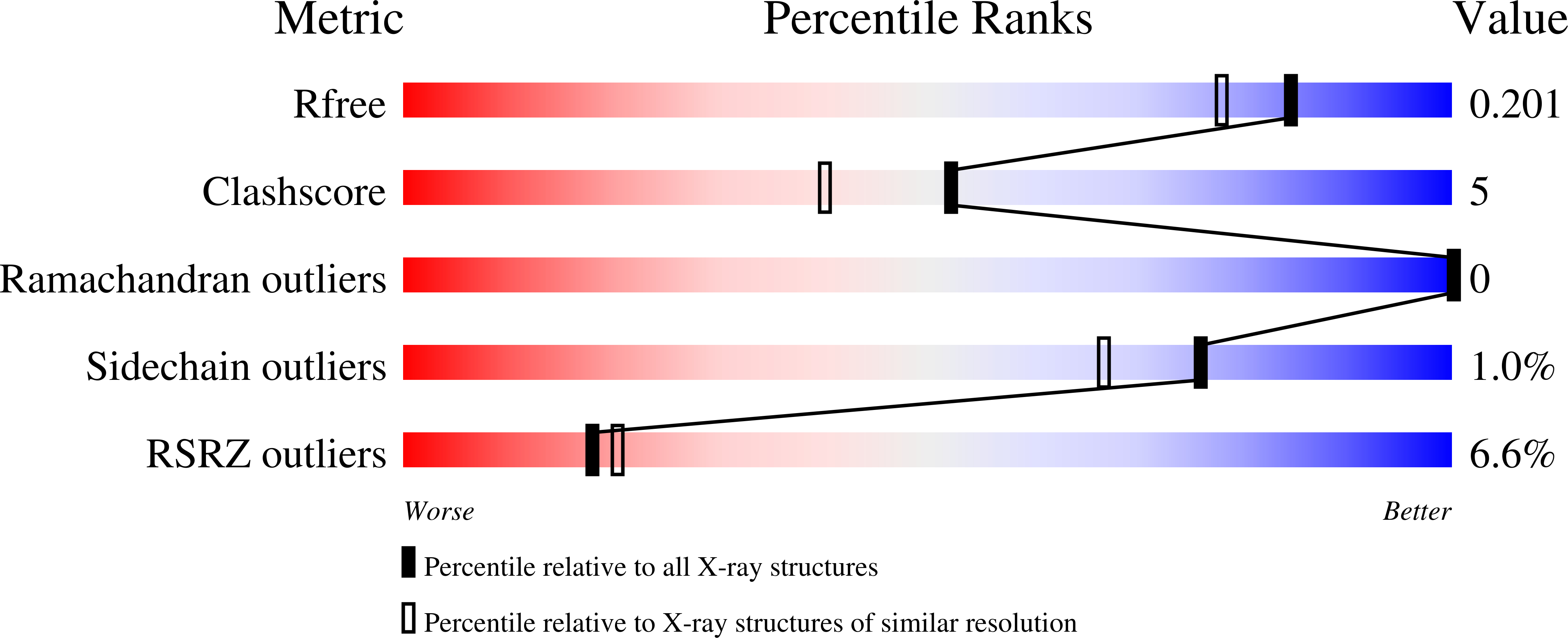
  • Method: X-RAY DIFFRACTION
  • Resolution: 1.70 Å
  • R-Value Free: 0.195 
  • R-Value Work: 0.166 
  • R-Value Observed: 0.168 

wwPDB Validation   3D Report Full Report


Ligand Structure Quality Assessment 


This is version 1.3 of the entry. See complete history


Literature

Structural basis of proteolytic activation of L-phenylalanine oxidase from Pseudomonas sp. P-501.

Ida, K.Kurabayashi, M.Suguro, M.Hiruma, Y.Hikima, T.Yamomoto, M.Suzuki, H.

(2008) J Biol Chem 283: 16584-16590

  • DOI: https://doi.org/10.1074/jbc.M800366200
  • Primary Citation of Related Structures:  
    2YR4, 2YR5, 2YR6

  • PubMed Abstract: 

    The mature form of l-phenylalanine oxidase (PAOpt) from Pseudomonas sp. P-501 was generated and activated by the proteolytic cleavage of a noncatalytic proenzyme (proPAO). The crystal structures of proPAO, PAOpt, and the PAOpt-o-amino benzoate (AB) complex were determined at 1.7, 1.25, and 1.35A resolutions, respectively. The structure of proPAO suggests that the prosequence peptide of proPAO occupies the funnel (pathway) of the substrate amino acid from the outside of the protein to the interior flavin ring, whereas the funnel is closed with the hydrophobic residues at its vestibule in both PAOpt and the PAOpt-AB complex. All three structures have an oxygen channel that is open to the surface of the protein from the flavin ring. These results suggest that structural changes were induced by proteolysis; that is, the proteolysis of proPAO removes the prosequence and closes the funnel to keep the active site hydrophobic but keeps the oxygen channel open. The possibility that an interaction of the hydrophobic side chain of substrate with the residues of the vestibule region may open the funnel as a putative amino acid channel is discussed.


  • Organizational Affiliation

    Department of Biosciences, School of Science., Graduate School of Fundamental Life Science, Kitasato University, Kitasato, Sagamihara, Kanagawa, Japan. idakoh@sci.kitasato-u.ac.jp


Macromolecules
Find similar proteins by:  (by identity cutoff)  |  3D Structure
Entity ID: 1
MoleculeChains Sequence LengthOrganismDetailsImage
Pro-enzyme of L-phenylalanine oxidase
A, B
721Pseudomonas sp. P-501Mutation(s): 0 
EC: 1.13.12.9
UniProt
Find proteins for Q5W9R9 (Pseudomonas sp)
Explore Q5W9R9 
Go to UniProtKB:  Q5W9R9
Entity Groups  
Sequence Clusters30% Identity50% Identity70% Identity90% Identity95% Identity100% Identity
UniProt GroupQ5W9R9
Sequence Annotations
Expand
  • Reference Sequence
Experimental Data & Validation

Experimental Data

  • Method: X-RAY DIFFRACTION
  • Resolution: 1.70 Å
  • R-Value Free: 0.195 
  • R-Value Work: 0.166 
  • R-Value Observed: 0.168 
  • Space Group: P 21 21 2
Unit Cell:
Length ( Å )Angle ( ˚ )
a = 141.577α = 90
b = 145.405β = 90
c = 82.076γ = 90
Software Package:
Software NamePurpose
REFMACrefinement
CNSrefinement
ADSCdata collection
MOSFLMdata reduction
SCALAdata scaling
SOLVEphasing

Structure Validation

View Full Validation Report


Ligand Structure Quality Assessment 


Entry History 

Deposition Data

Revision History  (Full details and data files)

  • Version 1.0: 2008-04-15
    Type: Initial release
  • Version 1.1: 2011-07-13
    Changes: Version format compliance
  • Version 1.2: 2012-12-12
    Changes: Database references
  • Version 1.3: 2024-03-13
    Changes: Data collection, Database references, Derived calculations