2ZNK

Thrombin Inhibition


Experimental Data Snapshot

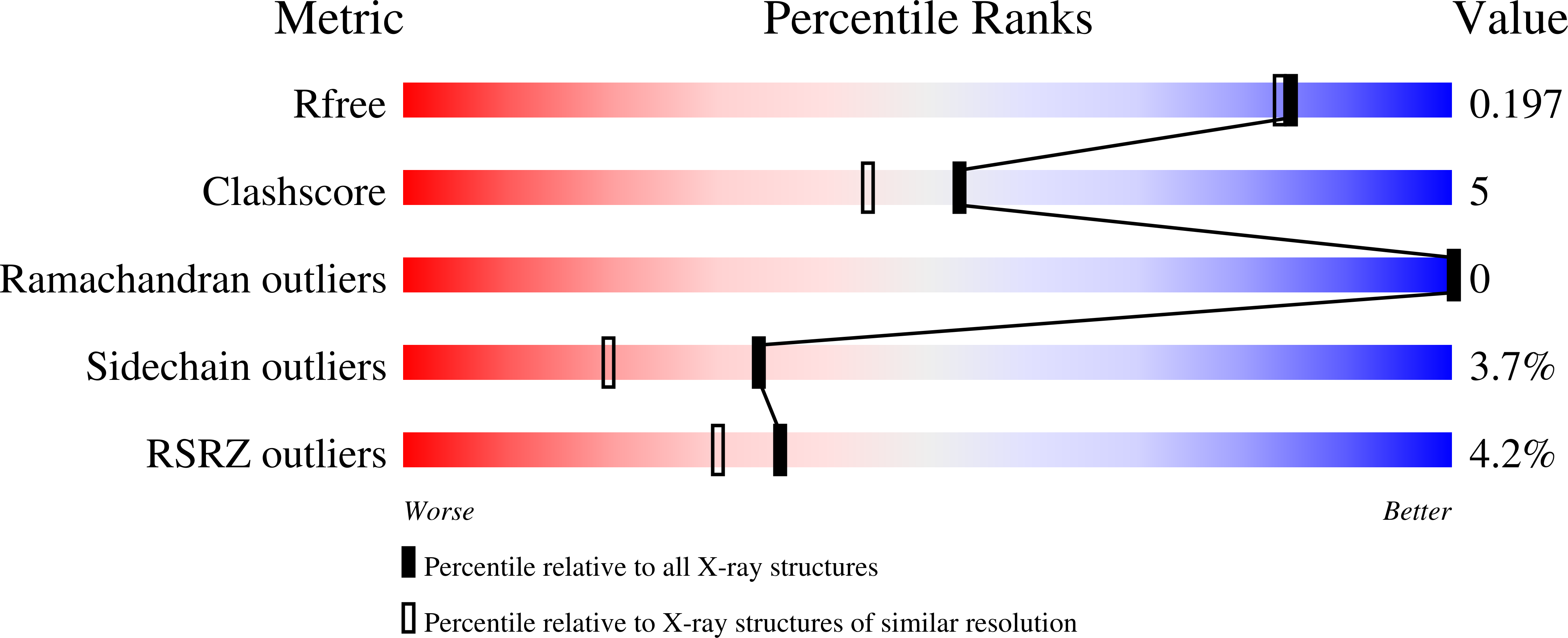
  • Method: X-RAY DIFFRACTION
  • Resolution: 1.80 Å
  • R-Value Free: 0.229 
  • R-Value Work: 0.175 
  • R-Value Observed: 0.180 

Starting Model: experimental
View more details

wwPDB Validation   3D Report Full Report


Ligand Structure Quality Assessment 


This is version 1.7 of the entry. See complete history


Literature

Non-additivity of functional group contributions in protein-ligand binding: a comprehensive study by crystallography and isothermal titration calorimetry.

Baum, B.Muley, L.Smolinski, M.Heine, A.Hangauer, D.Klebe, G.

(2010) J Mol Biol 397: 1042-1054

  • DOI: https://doi.org/10.1016/j.jmb.2010.02.007
  • Primary Citation of Related Structures:  
    2ZGB, 2ZHQ, 2ZI2, 2ZNK

  • PubMed Abstract: 

    Additivity of functional group contributions to protein-ligand binding is a very popular concept in medicinal chemistry as the basis of rational design and optimized lead structures. Most of the currently applied scoring functions for docking build on such additivity models. Even though the limitation of this concept is well known, case studies examining in detail why additivity fails at the molecular level are still very scarce. The present study shows, by use of crystal structure analysis and isothermal titration calorimetry for a congeneric series of thrombin inhibitors, that extensive cooperative effects between hydrophobic contacts and hydrogen bond formation are intimately coupled via dynamic properties of the formed complexes. The formation of optimal lipophilic contacts with the surface of the thrombin S3 pocket and the full desolvation of this pocket can conflict with the formation of an optimal hydrogen bond between ligand and protein. The mutual contributions of the competing interactions depend on the size of the ligand hydrophobic substituent and influence the residual mobility of ligand portions at the binding site. Analysis of the individual crystal structures and factorizing the free energy into enthalpy and entropy demonstrates that binding affinity of the ligands results from a mixture of enthalpic contributions from hydrogen bonding and hydrophobic contacts, and entropic considerations involving an increasing loss of residual mobility of the bound ligands. This complex picture of mutually competing and partially compensating enthalpic and entropic effects determines the non-additivity of free energy contributions to ligand binding at the molecular level.


  • Organizational Affiliation

    Department of Pharmaceutical Chemistry, Philipps-University Marburg, Marbacher Weg 6, 35032 Marburg, Germany.


Macromolecules
Find similar proteins by:  (by identity cutoff)  |  3D Structure
Entity ID: 1
MoleculeChains Sequence LengthOrganismDetailsImage
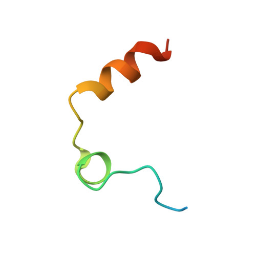
Thrombin light chainA [auth L]36Homo sapiensMutation(s): 0 
EC: 3.4.21.5
UniProt & NIH Common Fund Data Resources
Find proteins for P00734 (Homo sapiens)
Explore P00734 
Go to UniProtKB:  P00734
PHAROS:  P00734
GTEx:  ENSG00000180210 
Entity Groups  
Sequence Clusters30% Identity50% Identity70% Identity90% Identity95% Identity100% Identity
UniProt GroupP00734
Sequence Annotations
Expand
  • Reference Sequence
Find similar proteins by:  (by identity cutoff)  |  3D Structure
Entity ID: 2
MoleculeChains Sequence LengthOrganismDetailsImage
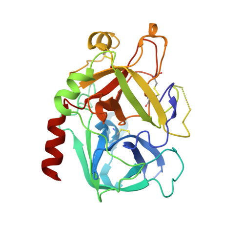
Thrombin heavy chainB [auth H]259Homo sapiensMutation(s): 0 
EC: 3.4.21.5
UniProt & NIH Common Fund Data Resources
Find proteins for P00734 (Homo sapiens)
Explore P00734 
Go to UniProtKB:  P00734
PHAROS:  P00734
GTEx:  ENSG00000180210 
Entity Groups  
Sequence Clusters30% Identity50% Identity70% Identity90% Identity95% Identity100% Identity
UniProt GroupP00734
Sequence Annotations
Expand
  • Reference Sequence

Find similar proteins by:  Sequence   |   3D Structure  

Entity ID: 3
MoleculeChains Sequence LengthOrganismDetailsImage
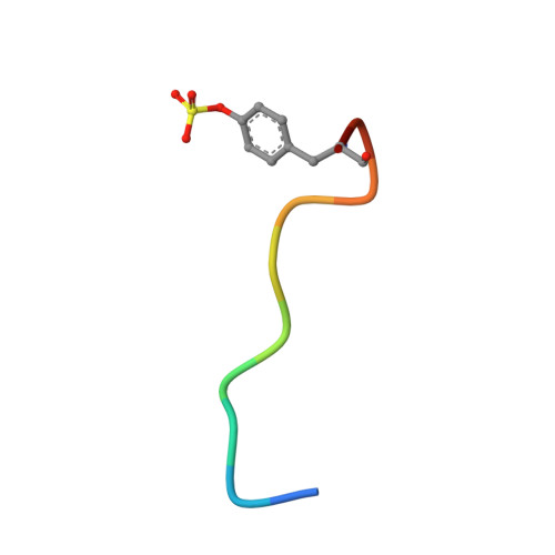
Hirudin variant-1C [auth I]11Hirudo medicinalisMutation(s): 0 
UniProt
Find proteins for P01050 (Hirudo medicinalis)
Explore P01050 
Go to UniProtKB:  P01050
Entity Groups  
Sequence Clusters30% Identity50% Identity70% Identity90% Identity95% Identity100% Identity
UniProt GroupP01050
Sequence Annotations
Expand
  • Reference Sequence
Small Molecules
Modified Residues  1 Unique
IDChains TypeFormula2D DiagramParent
TYS
Query on TYS
C [auth I]L-PEPTIDE LINKINGC9 H11 N O6 STYR
Biologically Interesting Molecules (External Reference) 1 Unique
Experimental Data & Validation

Experimental Data

  • Method: X-RAY DIFFRACTION
  • Resolution: 1.80 Å
  • R-Value Free: 0.229 
  • R-Value Work: 0.175 
  • R-Value Observed: 0.180 
  • Space Group: C 1 2 1
Unit Cell:
Length ( Å )Angle ( ˚ )
a = 70.2α = 90
b = 71.6β = 100.5
c = 72.5γ = 90
Software Package:
Software NamePurpose
SHELXmodel building
SHELXL-97refinement
MAR345data collection
HKL-2000data reduction
HKL-2000data scaling
SHELXphasing

Structure Validation

View Full Validation Report


Ligand Structure Quality Assessment 


Entry History 

Deposition Data

Revision History  (Full details and data files)

  • Version 1.0: 2009-04-14
    Type: Initial release
  • Version 1.1: 2011-07-13
    Changes: Atomic model, Database references, Derived calculations, Non-polymer description, Structure summary, Version format compliance
  • Version 1.2: 2012-12-12
    Changes: Other
  • Version 1.3: 2016-05-25
    Changes: Source and taxonomy
  • Version 1.4: 2017-10-11
    Changes: Refinement description
  • Version 1.5: 2023-11-01
    Changes: Data collection, Database references, Derived calculations, Refinement description
  • Version 1.6: 2023-11-15
    Changes: Data collection
  • Version 1.7: 2024-10-30
    Changes: Structure summary