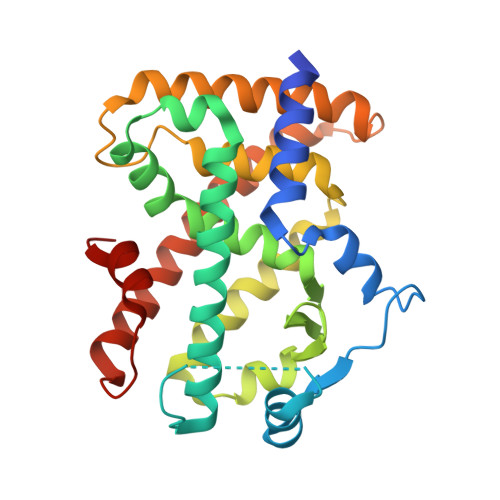
2ZNP

Human PPAR delta ligand binding domain in complex with a synthetic agonist TIPP204


Experimental Data Snapshot

  • Method: X-RAY DIFFRACTION
  • Resolution: 3.00 Å
  • R-Value Free: 0.288 
  • R-Value Work: 0.230 
  • R-Value Observed: 0.230 

Starting Model: experimental
View more details

wwPDB Validation   3D Report Full Report


Ligand Structure Quality Assessment 


This is version 1.5 of the entry. See complete history


Literature

Adaptability and selectivity of human peroxisome proliferator-activated receptor (PPAR) pan agonists revealed from crystal structures

Oyama, T.Toyota, K.Waku, T.Hirakawa, Y.Nagasawa, N.Kasuga, J.Hashimoto, Y.Miyachi, H.Morikawa, K.

(2009) Acta Crystallogr D Biol Crystallogr 65: 786-795

  • DOI: https://doi.org/10.1107/S0907444909015935
  • Primary Citation of Related Structures:  
    2ZNN, 2ZNO, 2ZNP, 2ZNQ

  • PubMed Abstract: 

    Peroxisome proliferator-activated receptors (PPARs) belong to the nuclear hormone receptor family, which is defined as transcriptional factors that are activated by the binding of ligands to their ligand-binding domains (LBDs). Although the three PPAR subtypes display different tissue distribution patterns and distinct pharmacological profiles, they all are essentially related to fatty-acid and glucose metabolism. Since the PPARs share similar three-dimensional structures within the LBDs, synthetic ligands which simultaneously activate two or all of the PPARs could be potent candidates in terms of drugs for the treatment of abnormal metabolic homeostasis. The structures of several PPAR LBDs were determined in complex with synthetic ligands, derivatives of 3-(4-alkoxyphenyl)propanoic acid, which exhibit unique agonistic activities. The PPARalpha and PPARgamma LBDs were complexed with the same pan agonist, TIPP-703, which activates all three PPARs and their crystal structures were determined. The two LBD-ligand complex structures revealed how the pan agonist is adapted to the similar, but significantly different, ligand-binding pockets of the PPARs. The structures of the PPARdelta LBD in complex with an alpha/delta-selective ligand, TIPP-401, and with a related delta-specific ligand, TIPP-204, were also determined. The comparison between the two PPARdelta complexes revealed how each ligand exhibits either a ;dual selective' or ;single specific' binding mode.


  • Organizational Affiliation

    The Takara Bio Endowed Division, Department of Biomolecular Recognition, Institute for Protein Research, Osaka University, Open Laboratories of Advanced Bioscience and Biotechnology, Suita, Osaka 565-0874, Japan.


Macromolecules
Find similar proteins by:  (by identity cutoff)  |  3D Structure
Entity ID: 1
MoleculeChains Sequence LengthOrganismDetailsImage
Peroxisome proliferator-activated receptor delta
A, B
276Homo sapiensMutation(s): 0 
Gene Names: PPARDNR1C2PPARB
UniProt & NIH Common Fund Data Resources
Find proteins for Q03181 (Homo sapiens)
Explore Q03181 
Go to UniProtKB:  Q03181
PHAROS:  Q03181
GTEx:  ENSG00000112033 
Entity Groups  
Sequence Clusters30% Identity50% Identity70% Identity90% Identity95% Identity100% Identity
UniProt GroupQ03181
Sequence Annotations
Expand
  • Reference Sequence
Small Molecules
Ligands 2 Unique
IDChains Name / Formula / InChI Key2D Diagram3D Interactions
K55
Query on K55

Download Ideal Coordinates CCD File 
D [auth A],
F [auth B]
(2S)-2-{4-butoxy-3-[({[2-fluoro-4-(trifluoromethyl)phenyl]carbonyl}amino)methyl]benzyl}butanoic acid
C24 H27 F4 N O4
AJSFKATVCYWKJN-INIZCTEOSA-N
B7G
Query on B7G

Download Ideal Coordinates CCD File 
C [auth A],
E [auth B]
heptyl beta-D-glucopyranoside
C13 H26 O6
NIDYWHLDTIVRJT-UJPOAAIJSA-N
Binding Affinity Annotations 
IDSourceBinding Affinity
K55 BindingDB:  2ZNP EC50: min: 0.72, max: 0.91 (nM) from 2 assay(s)
Experimental Data & Validation

Experimental Data

  • Method: X-RAY DIFFRACTION
  • Resolution: 3.00 Å
  • R-Value Free: 0.288 
  • R-Value Work: 0.230 
  • R-Value Observed: 0.230 
  • Space Group: P 1 21 1
Unit Cell:
Length ( Å )Angle ( ˚ )
a = 39.492α = 90
b = 93.149β = 97.48
c = 96.37γ = 90
Software Package:
Software NamePurpose
CNSrefinement
HKL-2000data collection
HKL-2000data reduction
HKL-2000data scaling
CNSphasing

Structure Validation

View Full Validation Report


Ligand Structure Quality Assessment 


Entry History 

Deposition Data

Revision History  (Full details and data files)

  • Version 1.0: 2009-05-05
    Type: Initial release
  • Version 1.1: 2011-07-13
    Changes: Version format compliance
  • Version 1.2: 2012-04-11
    Changes: Database references
  • Version 1.3: 2016-03-02
    Changes: Structure summary
  • Version 1.4: 2020-07-29
    Type: Remediation
    Reason: Carbohydrate remediation
    Changes: Data collection, Database references, Derived calculations, Structure summary
  • Version 1.5: 2023-11-01
    Changes: Data collection, Database references, Refinement description, Structure summary