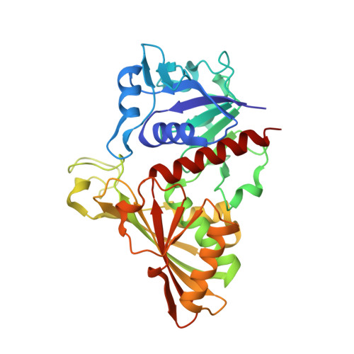
3B20 | pdb_00003b20

Crystal structure of Glyceraldehyde-3-Phosphate Dehydrogenase complexed with NADfrom Synechococcus elongatus"


Experimental Data Snapshot

  • Method: X-RAY DIFFRACTION
  • Resolution: 2.40 Å
  • R-Value Free: 
    0.269 (Depositor), 0.270 (DCC) 
  • R-Value Work: 
    0.213 (Depositor), 0.210 (DCC) 
  • R-Value Observed: 
    0.216 (Depositor) 

wwPDB Validation   3D Report Full Report


Ligand Structure Quality Assessment 

Created with Raphaël 2.3.0Worse 01 BetterLigand structure goodness of fit to experimental dataBest fitted NADClick on this verticalbar to view details

This is version 1.2 of the entry. See complete history


Literature

Structure Basis for the Regulation of Glyceraldehyde-3-Phosphate Dehydrogenase Activity via the Intrinsically Disordered Protein CP12.

Matsumura, H.Kai, A.Maeda, T.Tamoi, M.Satoh, A.Tamura, H.Hirose, M.Ogawa, T.Kizu, N.Wadano, A.Inoue, T.Shigeoka, S.

(2011) Structure 19: 1846-1854

  • DOI: https://doi.org/10.1016/j.str.2011.08.016
  • Primary Citation of Related Structures:  
    3B1J, 3B1K, 3B20

  • PubMed Abstract: 

    The reversible formation of a glyceraldehyde-3-phosphate dehydrogenase (GAPDH)-CP12-phosphoribulokinase (PRK) supramolecular complex, identified in oxygenic photosynthetic organisms, provides light-dependent Calvin cycle regulation in a coordinated manner. An intrinsically disordered protein (IDP) CP12 acts as a linker to sequentially bind GAPDH and PRK to downregulate both enzymes. Here, we report the crystal structures of the ternary GAPDH-CP12-NAD and binary GAPDH-NAD complexes from Synechococcus elongates. The GAPDH-CP12 complex structure reveals that the oxidized CP12 becomes partially structured upon GAPDH binding. The C-terminus of CP12 is inserted into the active-site region of GAPDH, resulting in competitive inhibition of GAPDH. This study also provides insight into how the GAPDH-CP12 complex is dissociated by a high NADP(H)/NAD(H) ratio. An unexpected increase in negative charge potential that emerged upon CP12 binding highlights the biological function of CP12 in the sequential assembly of the supramolecular complex.


  • Organizational Affiliation

    Department of Applied Chemistry, Graduate School of Engineering, Osaka University, 2-1 Yamadaoka, Suita, Osaka 565-0871, Japan. matsumura@chem.eng.osaka-u.ac.jp


Macromolecules
Find similar proteins by:  (by identity cutoff)  |  3D Structure
Entity ID: 1
MoleculeChains Sequence LengthOrganismDetailsImage
Glyceraldehyde 3-phosphate dehydrogenase (NADP+)339Synechococcus elongatus PCC 7942 = FACHB-805Mutation(s): 0 
Gene Names: gap2Synpcc7942_1742
EC: 1.2.1.13 (PDB Primary Data), 1.2.1 (UniProt)
UniProt
Find proteins for Q9R6W2 (Synechococcus elongatus (strain ATCC 33912 / PCC 7942 / FACHB-805))
Explore Q9R6W2 
Go to UniProtKB:  Q9R6W2
Entity Groups  
Sequence Clusters30% Identity50% Identity70% Identity90% Identity95% Identity100% Identity
UniProt GroupQ9R6W2
Sequence Annotations
Expand
  • Reference Sequence
Small Molecules
Ligands 2 Unique
IDChains Name / Formula / InChI Key2D Diagram3D Interactions
NAD
Query on NAD

Download Ideal Coordinates CCD File 
I [auth A]
L [auth B]
O
R [auth P]
U [auth Q]
NICOTINAMIDE-ADENINE-DINUCLEOTIDE
C21 H27 N7 O14 P2
BAWFJGJZGIEFAR-NNYOXOHSSA-N
SO4
Query on SO4

Download Ideal Coordinates CCD File 
G [auth A]
H [auth A]
J [auth B]
K [auth B]
M [auth O]
SULFATE ION
O4 S
QAOWNCQODCNURD-UHFFFAOYSA-L
Experimental Data & Validation

Experimental Data

  • Method: X-RAY DIFFRACTION
  • Resolution: 2.40 Å
  • R-Value Free:  0.269 (Depositor), 0.270 (DCC) 
  • R-Value Work:  0.213 (Depositor), 0.210 (DCC) 
  • R-Value Observed: 0.216 (Depositor) 
Space Group: C 1 2 1
Unit Cell:
Length ( Å )Angle ( ˚ )
a = 150.589α = 90
b = 79.847β = 101.65
c = 206.794γ = 90
Software Package:
Software NamePurpose
DNAdata collection
MOLREPphasing
PHENIXrefinement
HKL-2000data reduction
HKL-2000data scaling

Structure Validation

View Full Validation Report


Ligand Structure Quality Assessment 

Created with Raphaël 2.3.0Worse 01 BetterLigand structure goodness of fit to experimental dataBest fitted NADClick on this verticalbar to view details

Entry History 

Deposition Data

Revision History  (Full details and data files)

  • Version 1.0: 2012-01-11
    Type: Initial release
  • Version 1.1: 2013-06-12
    Changes: Structure summary
  • Version 1.2: 2024-03-13
    Changes: Data collection, Database references, Derived calculations