3BE1

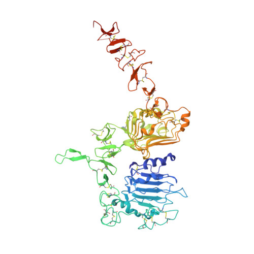
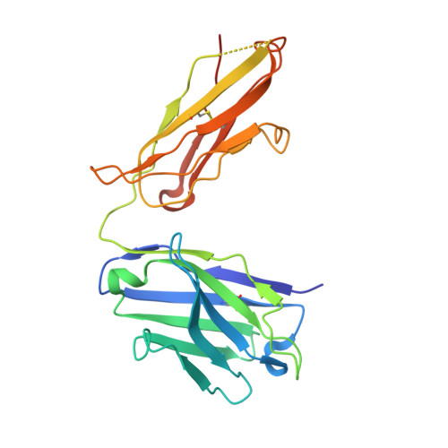
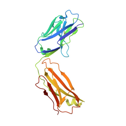
Dual specific bH1 Fab in complex with the extracellular domain of HER2/ErbB-2


Experimental Data Snapshot

  • Method: X-RAY DIFFRACTION
  • Resolution: 2.90 Å
  • R-Value Free: 0.277 
  • R-Value Work: 0.236 
  • R-Value Observed: 0.222 

Starting Model: experimental
View more details

wwPDB Validation   3D Report Full Report


Ligand Structure Quality Assessment 


This is version 1.5 of the entry. See complete history


Literature

Variants of the antibody herceptin that interact with HER2 and VEGF at the antigen binding site

Bostrom, J.Yu, S.F.Kan, D.Appleton, B.A.Lee, C.V.Billeci, K.Man, W.Peale, F.Ross, S.Wiesmann, C.Fuh, G.

(2009) Science 323: 1610-1614

  • DOI: https://doi.org/10.1126/science.1165480
  • Primary Citation of Related Structures:  
    3BDY, 3BE1

  • PubMed Abstract: 

    The interface between antibody and antigen is often depicted as a lock and key, suggesting that an antibody surface can accommodate only one antigen. Here, we describe an antibody with an antigen binding site that binds two distinct proteins with high affinity. We isolated a variant of Herceptin, a therapeutic monoclonal antibody that binds the human epidermal growth factor receptor 2 (HER2), on the basis of its ability to simultaneously interact with vascular endothelial growth factor (VEGF). Crystallographic and mutagenesis studies revealed that distinct amino acids of this antibody, called bH1, engage HER2 and VEGF energetically, but there is extensive overlap between the antibody surface areas contacting the two antigens. An affinity-improved version of bH1 inhibits both HER2- and VEGF-mediated cell proliferation in vitro and tumor progression in mouse models. Such "two-in-one" antibodies challenge the monoclonal antibody paradigm of one binding site, one antigen. They could also provide new opportunities for antibody-based therapy.


  • Organizational Affiliation

    Department of Protein Engineering, Genentech, 1 DNA Way, South San Francisco, CA 94080, USA.


Macromolecules
Find similar proteins by:  (by identity cutoff)  |  3D Structure
Entity ID: 1
MoleculeChains Sequence LengthOrganismDetailsImage
Receptor tyrosine-protein kinase erbB-2624Homo sapiensMutation(s): 0 
Gene Names: ERBB2HER2NEUNGL
EC: 2.7.10.1
UniProt & NIH Common Fund Data Resources
Find proteins for P04626 (Homo sapiens)
Explore P04626 
Go to UniProtKB:  P04626
PHAROS:  P04626
GTEx:  ENSG00000141736 
Entity Groups  
Sequence Clusters30% Identity50% Identity70% Identity90% Identity95% Identity100% Identity
UniProt GroupP04626
Glycosylation
Glycosylation Sites: 2Go to GlyGen: P04626-1
Sequence Annotations
Expand
  • Reference Sequence
Find similar proteins by:  (by identity cutoff)  |  3D Structure
Entity ID: 2
MoleculeChains Sequence LengthOrganismDetailsImage
Fab Fragment-Heavy ChainB [auth H]230Homo sapiensMutation(s): 0 
Entity Groups  
Sequence Clusters30% Identity50% Identity70% Identity90% Identity95% Identity100% Identity
Sequence Annotations
Expand
  • Reference Sequence
Find similar proteins by:  (by identity cutoff)  |  3D Structure
Entity ID: 3
MoleculeChains Sequence LengthOrganismDetailsImage
Fab Fragment-Light ChainC [auth L]218Homo sapiensMutation(s): 0 
Entity Groups  
Sequence Clusters30% Identity50% Identity70% Identity90% Identity95% Identity100% Identity
Sequence Annotations
Expand
  • Reference Sequence
Experimental Data & Validation

Experimental Data

  • Method: X-RAY DIFFRACTION
  • Resolution: 2.90 Å
  • R-Value Free: 0.277 
  • R-Value Work: 0.236 
  • R-Value Observed: 0.222 
  • Space Group: P 21 21 21
Unit Cell:
Length ( Å )Angle ( ˚ )
a = 62.324α = 90
b = 115.056β = 90
c = 208.171γ = 90
Software Package:
Software NamePurpose
DENZOdata reduction
SCALEPACKdata scaling
PHASERphasing
REFMACrefinement
PDB_EXTRACTdata extraction
HKL-2000data collection

Structure Validation

View Full Validation Report


Ligand Structure Quality Assessment 


Entry History 

Deposition Data

Revision History  (Full details and data files)

  • Version 1.0: 2008-11-18
    Type: Initial release
  • Version 1.1: 2011-07-13
    Changes: Advisory, Version format compliance
  • Version 1.2: 2017-10-25
    Changes: Refinement description
  • Version 1.3: 2020-07-29
    Type: Remediation
    Reason: Carbohydrate remediation
    Changes: Data collection, Derived calculations, Structure summary
  • Version 1.4: 2023-08-30
    Changes: Data collection, Database references, Refinement description, Structure summary
  • Version 1.5: 2024-11-13
    Changes: Structure summary