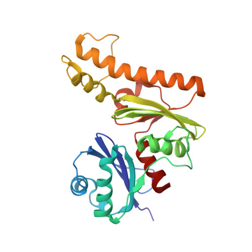
3BF1

Type III pantothenate kinase from Thermotoga maritima complexed with pantothenate and ADP


Experimental Data Snapshot

  • Method: X-RAY DIFFRACTION
  • Resolution: 2.30 Å
  • R-Value Free: 0.257 
  • R-Value Work: 0.190 
  • R-Value Observed: 0.194 

wwPDB Validation   3D Report Full Report


Ligand Structure Quality Assessment 


This is version 1.2 of the entry. See complete history


Literature

Structural basis for substrate binding and the catalytic mechanism of type III pantothenate kinase.

Yang, K.Strauss, E.Huerta, C.Zhang, H.

(2008) Biochemistry 47: 1369-1380

  • DOI: https://doi.org/10.1021/bi7018578
  • Primary Citation of Related Structures:  
    3BEX, 3BF1, 3BF3

  • PubMed Abstract: 

    Pantothenate kinase (PanK) catalyzes the first step of the universal five-step coenzyme A (CoA) biosynthetic pathway. The recently characterized type III PanK (PanK-III, encoded by the coaX gene) is distinct in sequence, structure and enzymatic properties from both the long-known bacterial type I PanK (PanK-I, exemplified by the Escherichia coli CoaA protein) and the predominantly eukaryotic type II PanK (PanK-II). PanK-III enzymes have an unusually high Km for ATP, are resistant to feedback inhibition by CoA, and are unable to utilize the N-alkylpantothenamide family of pantothenate analogues as alternative substrates, thus making type III PanK ineffective in generating CoA analogues as antimetabolites in vivo. Previously, we reported the crystal structure of the PanK-III from Thermotoga maritima and identified it as a member of the "acetate and sugar kinase/heat shock protein 70/actin" (ASKHA) superfamily. Here we report the crystal structures of the same PanK-III in complex with one of its substrates (pantothenate), its product (phosphopantothenate) as well as a ternary complex structure of PanK-III with pantothenate and ADP. These results are combined with isothermal titration calorimetry experiments to present a detailed structural and thermodynamic characterization of the interactions between PanK-III and its substrates ATP and pantothenate. Comparison of substrate binding and catalytic sites of PanK-III with that of eukaryotic PanK-II revealed drastic differences in the binding modes for both ATP and pantothenate substrates, and suggests that these differences may be exploited in the development of new inhibitors specifically targeting PanK-III.


  • Organizational Affiliation

    Department of Biochemistry, University of Texas Southwestern Medical Center, Dallas, Texas 75390, USA.


Macromolecules
Find similar proteins by:  (by identity cutoff)  |  3D Structure
Entity ID: 1
MoleculeChains Sequence LengthOrganismDetailsImage
Type III pantothenate kinase
A, B, C, D, E
A, B, C, D, E, F
249Thermotoga maritimaMutation(s): 0 
Gene Names: coaX
EC: 2.7.1.33
UniProt
Find proteins for Q9WZY5 (Thermotoga maritima (strain ATCC 43589 / DSM 3109 / JCM 10099 / NBRC 100826 / MSB8))
Explore Q9WZY5 
Go to UniProtKB:  Q9WZY5
Entity Groups  
Sequence Clusters30% Identity50% Identity70% Identity90% Identity95% Identity100% Identity
UniProt GroupQ9WZY5
Sequence Annotations
Expand
  • Reference Sequence
Small Molecules
Ligands 2 Unique
IDChains Name / Formula / InChI Key2D Diagram3D Interactions
ADP
Query on ADP

Download Ideal Coordinates CCD File 
G [auth A]
I [auth B]
K [auth C]
M [auth D]
O [auth E]
G [auth A],
I [auth B],
K [auth C],
M [auth D],
O [auth E],
Q [auth F]
ADENOSINE-5'-DIPHOSPHATE
C10 H15 N5 O10 P2
XTWYTFMLZFPYCI-KQYNXXCUSA-N
PAU
Query on PAU

Download Ideal Coordinates CCD File 
H [auth A]
J [auth B]
L [auth C]
N [auth D]
P [auth E]
H [auth A],
J [auth B],
L [auth C],
N [auth D],
P [auth E],
R [auth F]
PANTOTHENOIC ACID
C9 H17 N O5
GHOKWGTUZJEAQD-ZETCQYMHSA-N
Binding Affinity Annotations 
IDSourceBinding Affinity
ADP PDBBind:  3BF1 Kd: 3.00e+6 (nM) from 1 assay(s)
Experimental Data & Validation

Experimental Data

  • Method: X-RAY DIFFRACTION
  • Resolution: 2.30 Å
  • R-Value Free: 0.257 
  • R-Value Work: 0.190 
  • R-Value Observed: 0.194 
  • Space Group: P 1 21 1
Unit Cell:
Length ( Å )Angle ( ˚ )
a = 74.962α = 90
b = 136.685β = 109.71
c = 75.01γ = 90
Software Package:
Software NamePurpose
REFMACrefinement
HKL-2000data collection
HKL-2000data reduction
HKL-2000data scaling

Structure Validation

View Full Validation Report


Ligand Structure Quality Assessment 


Entry History 

Deposition Data

Revision History  (Full details and data files)

  • Version 1.0: 2008-06-24
    Type: Initial release
  • Version 1.1: 2011-07-13
    Changes: Version format compliance
  • Version 1.2: 2024-02-21
    Changes: Data collection, Database references, Derived calculations, Refinement description