3D7W | pdb_00003d7w

Mistletoe Lectin I in Complex with Zeatin


Experimental Data Snapshot

  • Method: X-RAY DIFFRACTION
  • Resolution: 2.49 Å
  • R-Value Free: 
    0.224 (Depositor), 0.220 (DCC) 
  • R-Value Work: 
    0.182 (Depositor), 0.180 (DCC) 
  • R-Value Observed: 
    0.184 (Depositor) 

Starting Model: experimental
View more details

wwPDB Validation   3D Report Full Report


Ligand Structure Quality Assessment 

Created with Raphaël 2.3.0Worse 01 BetterLigand structure goodness of fit to experimental dataBest fitted NAGClick on this verticalbar to view detailsBest fitted ZEZClick on this verticalbar to view detailsBest fitted ZEAClick on this verticalbar to view details

This is version 2.2 of the entry. See complete history


Literature

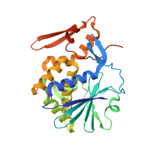
Structure of mistletoe lectin I from Viscum album in complex with the phytohormone zeatin

Meyer, A.Rypniewski, W.Szymanski, M.Voelter, W.Barciszewski, J.Betzel, C.

(2008) Biochim Biophys Acta 1784: 1590-1595

  • DOI: https://doi.org/10.1016/j.bbapap.2008.07.010
  • Primary Citation of Related Structures:  
    3D7W

  • PubMed Abstract: 

    The crystal structure of mistletoe lectin I (ML-I) isolated from the European mistletoe Viscum album in complex with the most active phytohormone zeatin has been analyzed and refined to 2.54 A resolution. X-ray suitable crystals of ML-I were obtained by the counter-diffusion method using the Gel-Tube R crystallization kit (GT-R) onboard the Russian Service Module on the international space station ISS. High quality hexagonal bipyramidal crystals were grown during 3 months under microgravity conditions. Selected crystals were soaked in a saturated solution of zeatin and subsequently diffraction data were collected applying synchrotron radiation. A distinct F(o)-F(c) electron density has been found inside a binding pocket located in subunit B of ML-I and has been interpreted as a single zeatin molecule. The structure was refined to investigate the zeatin-ML-I interactions in detail. The results demonstrate the ability of mistletoe to protect itself from the host transpiration regulation by absorbing the most active host plant hormones as part of a defense mechanism.


  • Organizational Affiliation

    Institute of Biochemistry and Molecular Biology, University of Hamburg, c/o DESY, Notkestr. 85, Building 22a, 22603 Hamburg, Germany.


Macromolecules
Find similar proteins by:  (by identity cutoff)  |  3D Structure
Entity ID: 1
MoleculeChains Sequence LengthOrganismDetailsImage
Beta-galactoside-specific lectin 1254Viscum albumMutation(s): 0 
EC: 3.2.2.22
UniProt
Find proteins for P81446 (Viscum album)
Explore P81446 
Go to UniProtKB:  P81446
Entity Groups  
Sequence Clusters30% Identity50% Identity70% Identity90% Identity95% Identity100% Identity
UniProt GroupP81446
Glycosylation
Glycosylation Sites: 1
Sequence Annotations
Expand
  • Reference Sequence
Find similar proteins by:  (by identity cutoff)  |  3D Structure
Entity ID: 2
MoleculeChains Sequence LengthOrganismDetailsImage
Beta-galactoside-specific lectin 1265Viscum albumMutation(s): 0 
EC: 3.2.2.22
UniProt
Find proteins for P81446 (Viscum album)
Explore P81446 
Go to UniProtKB:  P81446
Entity Groups  
Sequence Clusters30% Identity50% Identity70% Identity90% Identity95% Identity100% Identity
UniProt GroupP81446
Glycosylation
Glycosylation Sites: 3
Sequence Annotations
Expand
  • Reference Sequence
Oligosaccharides

Help

Entity ID: 3
MoleculeChains Length2D Diagram Glycosylation3D Interactions
2-acetamido-2-deoxy-beta-D-glucopyranose-(1-4)-2-acetamido-2-deoxy-beta-D-glucopyranose
C, D
2N-Glycosylation
Glycosylation Resources
GlyTouCan:  G42666HT
GlyCosmos:  G42666HT
GlyGen:  G42666HT
Small Molecules
Ligands 5 Unique
IDChains Name / Formula / InChI Key2D Diagram3D Interactions
NAG
Query on NAG

Download Ideal Coordinates CCD File 
E [auth A],
J [auth B]
2-acetamido-2-deoxy-beta-D-glucopyranose
C8 H15 N O6
OVRNDRQMDRJTHS-FMDGEEDCSA-N
ZEZ
Query on ZEZ

Download Ideal Coordinates CCD File 
L [auth B](2Z)-2-methyl-4-(9H-purin-6-ylamino)but-2-en-1-ol
C10 H13 N5 O
UZKQTCBAMSWPJD-UQCOIBPSSA-N
ZEA
Query on ZEA

Download Ideal Coordinates CCD File 
K [auth B](2E)-2-methyl-4-(9H-purin-6-ylamino)but-2-en-1-ol
C10 H13 N5 O
UZKQTCBAMSWPJD-FARCUNLSSA-N
SO4
Query on SO4

Download Ideal Coordinates CCD File 
F [auth A],
G [auth A]
SULFATE ION
O4 S
QAOWNCQODCNURD-UHFFFAOYSA-L
GOL
Query on GOL

Download Ideal Coordinates CCD File 
H [auth A],
I [auth A],
M [auth B],
N [auth B]
GLYCEROL
C3 H8 O3
PEDCQBHIVMGVHV-UHFFFAOYSA-N
Experimental Data & Validation

Experimental Data

  • Method: X-RAY DIFFRACTION
  • Resolution: 2.49 Å
  • R-Value Free:  0.224 (Depositor), 0.220 (DCC) 
  • R-Value Work:  0.182 (Depositor), 0.180 (DCC) 
  • R-Value Observed: 0.184 (Depositor) 
Space Group: P 65 2 2
Unit Cell:
Length ( Å )Angle ( ˚ )
a = 106.644α = 90
b = 106.644β = 90
c = 311.667γ = 120
Software Package:
Software NamePurpose
REFMACrefinement

Structure Validation

View Full Validation Report


Ligand Structure Quality Assessment 

Created with Raphaël 2.3.0Worse 01 BetterLigand structure goodness of fit to experimental dataBest fitted NAGClick on this verticalbar to view detailsBest fitted ZEZClick on this verticalbar to view detailsBest fitted ZEAClick on this verticalbar to view details

Entry History 

Deposition Data

Revision History  (Full details and data files)

  • Version 1.0: 2008-06-17
    Type: Initial release
  • Version 1.1: 2011-07-13
    Changes: Non-polymer description, Version format compliance
  • Version 1.2: 2014-02-05
    Changes: Derived calculations
  • Version 2.0: 2020-07-29
    Type: Remediation
    Reason: Carbohydrate remediation
    Changes: Advisory, Atomic model, Data collection, Derived calculations, Structure summary
  • Version 2.1: 2023-11-01
    Changes: Data collection, Database references, Refinement description, Structure summary
  • Version 2.2: 2024-10-30
    Changes: Structure summary