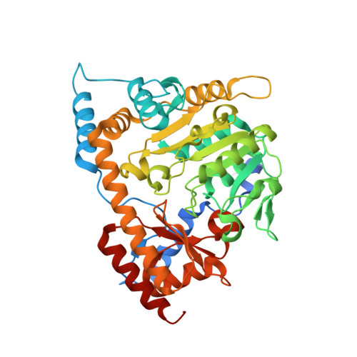
3EI8

Crystal structure of K270N variant of LL-diaminopimelate aminotransferase from Arabidopsis thaliana complexed with LL-DAP: External aldimine form


Experimental Data Snapshot

  • Method: X-RAY DIFFRACTION
  • Resolution: 1.60 Å
  • R-Value Free: 0.188 
  • R-Value Work: 0.167 
  • R-Value Observed: 0.168 

Starting Model: experimental
View more details

wwPDB Validation   3D Report Full Report


Ligand Structure Quality Assessment 


This is version 1.3 of the entry. See complete history


Literature

Mechanism of substrate recognition and PLP-induced conformational changes in LL-diaminopimelate aminotransferase from Arabidopsis thaliana.

Watanabe, N.Clay, M.D.van Belkum, M.J.Cherney, M.M.Vederas, J.C.James, M.N.

(2008) J Mol Biol 384: 1314-1329

  • DOI: https://doi.org/10.1016/j.jmb.2008.10.022
  • Primary Citation of Related Structures:  
    3EI5, 3EI6, 3EI7, 3EI8, 3EI9, 3EIA, 3EIB

  • PubMed Abstract: 

    LL-Diaminopimelate aminotransferase (LL-DAP-AT), a pyridoxal phosphate (PLP)-dependent enzyme in the lysine biosynthetic pathways of plants and Chlamydia, is a potential target for the development of herbicides or antibiotics. This homodimeric enzyme converts L-tetrahydrodipicolinic acid (THDP) directly to LL-DAP using L-glutamate as the source of the amino group. Earlier, we described the 3D structures of native and malate-bound LL-DAP-AT from Arabidopsis thaliana (AtDAP-AT). Seven additional crystal structures of AtDAP-AT and its variants are reported here as part of an investigation into the mechanism of substrate recognition and catalysis. Two structures are of AtDAP-AT with reduced external aldimine analogues: N-(5'-phosphopyridoxyl)-L-glutamate (PLP-Glu) and N-(5'-phosphopyridoxyl)- LL-Diaminopimelate (PLP-DAP) bound in the active site. Surprisingly, they reveal that both L-glutamate and LL-DAP are recognized in a very similar fashion by the same sets of amino acid residues; both molecules adopt twisted V-shaped conformations. With both substrates, the alpha-carboxylates are bound in a salt bridge with Arg404, whereas the distal carboxylates are recognized via hydrogen bonds to the well-conserved side chains of Tyr37, Tyr125 and Lys129. The distal C(epsilon) amino group of LL-DAP is specifically recognized by several non-covalent interactions with residues from the other subunit (Asn309*, Tyr94*, Gly95*, and Glu97* (Amino acid designators followed by an asterisk (*) indicate that the residues originate in the other subunit of the dimer)) and by three bound water molecules. Two catalytically inactive variants of AtDAP-AT were created via site-directed mutagenesis of the active site lysine (K270N and K270Q). The structures of these variants permitted the observation of the unreduced external aldimines of PLP with L-glutamate and with LL-DAP in the active site, and revealed differences in the torsion angle about the PLP-substrate bond. Lastly, an apo-AtDAP-AT structure missing PLP revealed details of conformational changes induced by PLP binding and substrate entry into the active site.


  • Organizational Affiliation

    Department of Biochemistry, University of Alberta, Edmonton, Alberta, Canada T6G 2H7.


Macromolecules
Find similar proteins by:  (by identity cutoff)  |  3D Structure
Entity ID: 1
MoleculeChains Sequence LengthOrganismDetailsImage
LL-diaminopimelate aminotransferase
A, B
432Arabidopsis thalianaMutation(s): 1 
Gene Names: DAPAGD2At4g33680T16L1.170
EC: 2.6.1.83
UniProt
Find proteins for Q93ZN9 (Arabidopsis thaliana)
Explore Q93ZN9 
Go to UniProtKB:  Q93ZN9
Entity Groups  
Sequence Clusters30% Identity50% Identity70% Identity90% Identity95% Identity100% Identity
UniProt GroupQ93ZN9
Sequence Annotations
Expand
  • Reference Sequence
Small Molecules
Ligands 4 Unique
IDChains Name / Formula / InChI Key2D Diagram3D Interactions
PL5
Query on PL5

Download Ideal Coordinates CCD File 
H [auth B](2S,6S)-2-amino-6-{[(1E)-{3-hydroxy-2-methyl-5-[(phosphonooxy)methyl]pyridin-4-yl}methylidene]amino}heptanedioic acid
C15 H22 N3 O9 P
VHNMKXLQZMEZSG-NTQSYZAISA-N
PLP
Query on PLP

Download Ideal Coordinates CCD File 
C [auth A]PYRIDOXAL-5'-PHOSPHATE
C8 H10 N O6 P
NGVDGCNFYWLIFO-UHFFFAOYSA-N
SO4
Query on SO4

Download Ideal Coordinates CCD File 
D [auth A],
I [auth B]
SULFATE ION
O4 S
QAOWNCQODCNURD-UHFFFAOYSA-L
GOL
Query on GOL

Download Ideal Coordinates CCD File 
E [auth A]
F [auth A]
G [auth A]
J [auth B]
K [auth B]
E [auth A],
F [auth A],
G [auth A],
J [auth B],
K [auth B],
L [auth B]
GLYCEROL
C3 H8 O3
PEDCQBHIVMGVHV-UHFFFAOYSA-N
Experimental Data & Validation

Experimental Data

  • Method: X-RAY DIFFRACTION
  • Resolution: 1.60 Å
  • R-Value Free: 0.188 
  • R-Value Work: 0.167 
  • R-Value Observed: 0.168 
  • Space Group: P 32 2 1
Unit Cell:
Length ( Å )Angle ( ˚ )
a = 102.476α = 90
b = 102.476β = 90
c = 171.68γ = 120
Software Package:
Software NamePurpose
Blu-Icedata collection
MOLREPphasing
REFMACrefinement
HKL-2000data reduction
HKL-2000data scaling

Structure Validation

View Full Validation Report


Ligand Structure Quality Assessment 


Entry History 

Deposition Data

Revision History  (Full details and data files)

  • Version 1.0: 2008-10-14
    Type: Initial release
  • Version 1.1: 2011-07-13
    Changes: Non-polymer description, Version format compliance
  • Version 1.2: 2021-10-20
    Changes: Database references, Derived calculations
  • Version 1.3: 2023-08-30
    Changes: Data collection, Refinement description