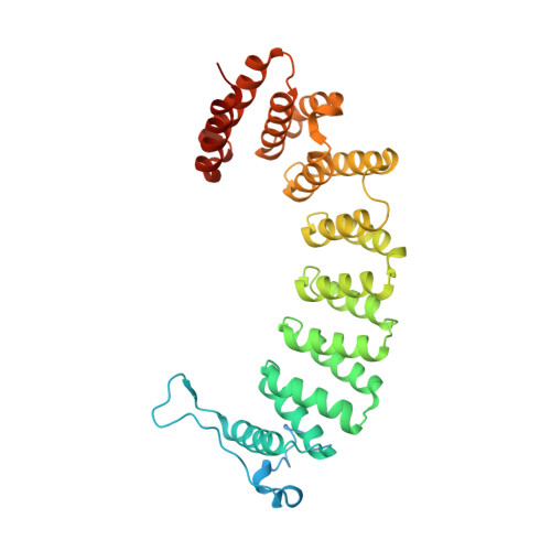
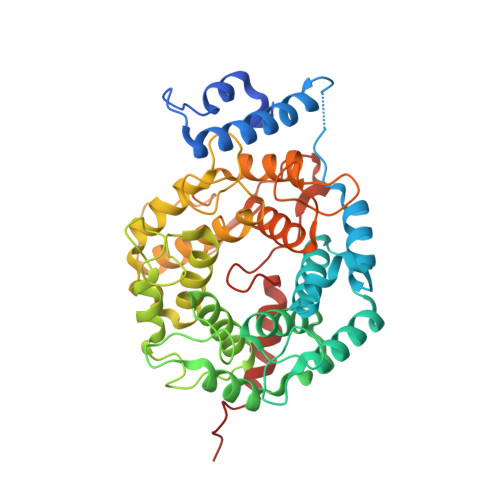
3EU5

Crystal structure of FTase(ALPHA-subunit; BETA-subunit DELTA C10) in complex with BiotinGPP


Experimental Data Snapshot

  • Method: X-RAY DIFFRACTION
  • Resolution: 2.80 Å
  • R-Value Free: 0.207 
  • R-Value Work: 0.153 
  • R-Value Observed: 0.155 

Starting Model: experimental
View more details

wwPDB Validation   3D Report Full Report


Ligand Structure Quality Assessment 


This is version 1.2 of the entry. See complete history


Literature

Analysis of the eukaryotic prenylome by isoprenoid affinity tagging

Nguyen, U.T.T.Guo, Z.Delon, C.Wu, Y.Deraeve, C.Franzel, B.Bon, R.S.Blankenfeldt, W.Goody, R.S.Waldmann, H.Wolters, D.Alexandrov, K.

(2009) Nat Chem Biol 5: 227-235

  • DOI: https://doi.org/10.1038/nchembio.149
  • Primary Citation of Related Structures:  
    3EU5, 3EUV

  • PubMed Abstract: 

    Protein prenylation is a widespread phenomenon in eukaryotic cells that affects many important signaling molecules. We describe the structure-guided design of engineered protein prenyltransferases and their universal synthetic substrate, biotin-geranylpyrophosphate. These new tools allowed us to detect femtomolar amounts of prenylatable proteins in cells and organs and to identify their cognate protein prenyltransferases. Using this approach, we analyzed the in vivo effects of protein prenyltransferase inhibitors. Whereas some of the inhibitors displayed the expected activities, others lacked in vivo activity or targeted a broader spectrum of prenyltransferases than previously believed. To quantitate the in vivo effect of the prenylation inhibitors, we profiled biotin-geranyl-tagged RabGTPases across the proteome by mass spectrometry. We also demonstrate that sites of active vesicular transport carry most of the RabGTPases. This approach enables a quantitative proteome-wide analysis of the regulation of protein prenylation and its modulation by therapeutic agents.


  • Organizational Affiliation

    Max Planck Institute of Molecular Physiology, Dortmund, Germany.


Macromolecules
Find similar proteins by:  (by identity cutoff)  |  3D Structure
Entity ID: 1
MoleculeChains Sequence LengthOrganismDetailsImage
Protein farnesyltransferase/geranylgeranyltransferase type-1 subunit alpha377Rattus norvegicusMutation(s): 0 
EC: 2.5.1.58 (PDB Primary Data), 2.5.1.59 (PDB Primary Data)
UniProt
Find proteins for Q04631 (Rattus norvegicus)
Explore Q04631 
Go to UniProtKB:  Q04631
Entity Groups  
Sequence Clusters30% Identity50% Identity70% Identity90% Identity95% Identity100% Identity
UniProt GroupQ04631
Sequence Annotations
Expand
  • Reference Sequence
Find similar proteins by:  (by identity cutoff)  |  3D Structure
Entity ID: 2
MoleculeChains Sequence LengthOrganismDetailsImage
Protein farnesyltransferase subunit beta427Rattus norvegicusMutation(s): 0 
EC: 2.5.1.58
UniProt
Find proteins for Q02293 (Rattus norvegicus)
Explore Q02293 
Go to UniProtKB:  Q02293
Entity Groups  
Sequence Clusters30% Identity50% Identity70% Identity90% Identity95% Identity100% Identity
UniProt GroupQ02293
Sequence Annotations
Expand
  • Reference Sequence
Small Molecules
Ligands 2 Unique
IDChains Name / Formula / InChI Key2D Diagram3D Interactions
GBO
Query on GBO

Download Ideal Coordinates CCD File 
D [auth B](2E,6E)-3,7-dimethyl-8-({5-[(3aS,4S,6aR)-2-oxohexahydro-1H-thieno[3,4-d]imidazol-4-yl]pentanoyl}amino)octa-2,6-dien-1-yl trihydrogen diphosphate
C20 H35 N3 O9 P2 S
XZJFCDBLIMGOSX-VRLLXNLQSA-N
ZN
Query on ZN

Download Ideal Coordinates CCD File 
C [auth B]ZINC ION
Zn
PTFCDOFLOPIGGS-UHFFFAOYSA-N
Binding Affinity Annotations 
IDSourceBinding Affinity
GBO PDBBind:  3EU5 Kd: 32 (nM) from 1 assay(s)
Experimental Data & Validation

Experimental Data

  • Method: X-RAY DIFFRACTION
  • Resolution: 2.80 Å
  • R-Value Free: 0.207 
  • R-Value Work: 0.153 
  • R-Value Observed: 0.155 
  • Space Group: P 61
Unit Cell:
Length ( Å )Angle ( ˚ )
a = 175.065α = 90
b = 175.065β = 90
c = 70.618γ = 120
Software Package:
Software NamePurpose
PHASERphasing
REFMACrefinement
XDSdata reduction
XSCALEdata scaling

Structure Validation

View Full Validation Report


Ligand Structure Quality Assessment 


Entry History 

Deposition Data

Revision History  (Full details and data files)

  • Version 1.0: 2009-07-07
    Type: Initial release
  • Version 1.1: 2011-07-13
    Changes: Version format compliance
  • Version 1.2: 2023-11-01
    Changes: Data collection, Database references, Derived calculations, Refinement description