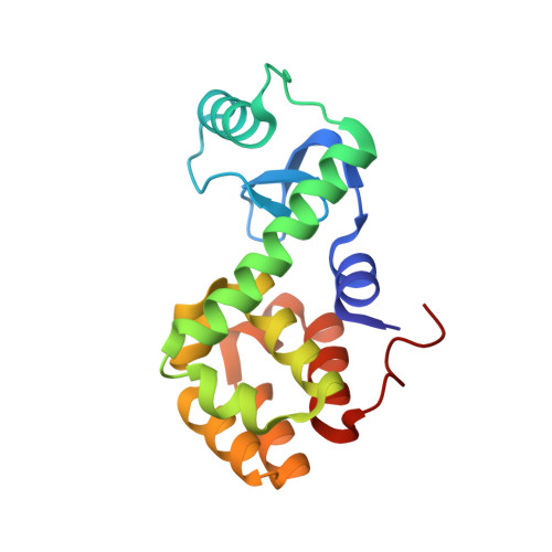
3F8V

Evaulaution at Atomic Resolution of the Role of Strain in Destabilizing the Temperature Sensitive T4 Lysozyme Mutant Arg96-->His


Experimental Data Snapshot

  • Method: X-RAY DIFFRACTION
  • Resolution: 1.08 Å
  • R-Value Free: 0.163 
  • R-Value Work: 0.129 
  • R-Value Observed: 0.138 

Starting Model: experimental
View more details

wwPDB Validation   3D Report Full Report


Ligand Structure Quality Assessment 


This is version 1.5 of the entry. See complete history


Literature

Evaluation at atomic resolution of the role of strain in destabilizing the temperature-sensitive T4 lysozyme mutant Arg 96 --> His.

Mooers, B.H.Tronrud, D.E.Matthews, B.W.

(2009) Protein Sci 18: 863-870

  • DOI: https://doi.org/10.1002/pro.93
  • Primary Citation of Related Structures:  
    3F8V, 3F9L, 3FA0, 3FAD

  • PubMed Abstract: 

    Mutant R96H is a classic temperature-sensitive mutant of bacteriophage T4 lysozyme. It was in fact the first variant of the protein to be characterized structurally. Subsequently, it has been studied extensively by a variety of experimental and computational techniques, but the reasons for the loss of stability of the mutant protein remain controversial. In the crystallographic refinement of the mutant structure at 1.9 A resolution one of the bond angles at the site of substitution appeared to be distorted by about 11( degrees ), and it was suggested that this steric strain was one of the major factors in destabilizing the mutant. Different computationally-derived models of the mutant structure, however, did not show such distortion. To determine the geometry at the site of mutation more reliably, we have extended the resolution of the data and refined the wildtype (WT) and mutant structures to be better than 1.1 A resolution. The high-resolution refinement of the structure of R96H does not support the bond angle distortion seen in the 1.9 A structure determination. At the same time, it does confirm other manifestations of strain seen previously including an unusual rotameric state for His96 with distorted hydrogen bonding. The rotamer strain has been estimated as about 0.8 kcal/mol, which is about 25% of the overall reduction in stability of the mutant. Because of concern that contacts from a neighboring molecule in the crystal might influence the geometry at the site of mutation we also constructed and analyzed supplemental mutant structures in which this crystal contact was eliminated. High-resolution refinement shows that the crystal contacts have essentially no effect on the conformation of Arg96 in WT or on His96 in the R96H mutant.


  • Organizational Affiliation

    Howard Hughes Medical Institute, Institute of Molecular Biology and Department of Physics, 1229 University of Oregon Eugene, Oregon 97403-1229, USA.


Macromolecules
Find similar proteins by:  (by identity cutoff)  |  3D Structure
Entity ID: 1
MoleculeChains Sequence LengthOrganismDetailsImage
Lysozyme164Tequatrovirus T4Mutation(s): 1 
Gene Names: gene e
EC: 3.2.1.17
UniProt
Find proteins for P00720 (Enterobacteria phage T4)
Explore P00720 
Go to UniProtKB:  P00720
Entity Groups  
Sequence Clusters30% Identity50% Identity70% Identity90% Identity95% Identity100% Identity
UniProt GroupP00720
Sequence Annotations
Expand
  • Reference Sequence
Small Molecules
Ligands 5 Unique
IDChains Name / Formula / InChI Key2D Diagram3D Interactions
HED
Query on HED

Download Ideal Coordinates CCD File 
B [auth A]2-HYDROXYETHYL DISULFIDE
C4 H10 O2 S2
KYNFOMQIXZUKRK-UHFFFAOYSA-N
PO4
Query on PO4

Download Ideal Coordinates CCD File 
C [auth A]PHOSPHATE ION
O4 P
NBIIXXVUZAFLBC-UHFFFAOYSA-K
K
Query on K

Download Ideal Coordinates CCD File 
G [auth A]POTASSIUM ION
K
NPYPAHLBTDXSSS-UHFFFAOYSA-N
CL
Query on CL

Download Ideal Coordinates CCD File 
D [auth A],
F [auth A]
CHLORIDE ION
Cl
VEXZGXHMUGYJMC-UHFFFAOYSA-M
NA
Query on NA

Download Ideal Coordinates CCD File 
E [auth A]SODIUM ION
Na
FKNQFGJONOIPTF-UHFFFAOYSA-N
Experimental Data & Validation

Experimental Data

  • Method: X-RAY DIFFRACTION
  • Resolution: 1.08 Å
  • R-Value Free: 0.163 
  • R-Value Work: 0.129 
  • R-Value Observed: 0.138 
  • Space Group: P 32 2 1
Unit Cell:
Length ( Å )Angle ( ˚ )
a = 60.123α = 90
b = 60.123β = 90
c = 95.445γ = 120
Software Package:
Software NamePurpose
MAR345dtbdata collection
SHELXmodel building
SHELXL-97refinement
MOSFLMdata reduction
SCALAdata scaling
SHELXphasing

Structure Validation

View Full Validation Report


Ligand Structure Quality Assessment 


Entry History 

Deposition Data

Revision History  (Full details and data files)

  • Version 1.0: 2009-02-17
    Type: Initial release
  • Version 1.1: 2011-07-13
    Changes: Version format compliance
  • Version 1.2: 2018-01-24
    Changes: Structure summary
  • Version 1.3: 2021-10-20
    Changes: Database references, Derived calculations
  • Version 1.4: 2023-12-27
    Changes: Data collection
  • Version 1.5: 2024-04-03
    Changes: Refinement description