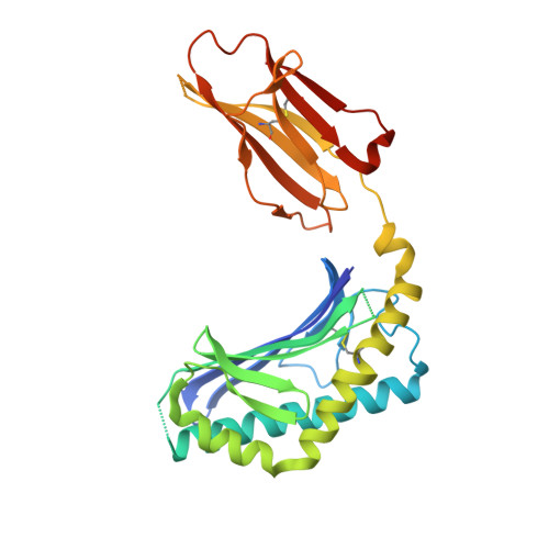
3G08

Crystal structure of the alpha-galactosylceramide analog OCH in complex with mouse CD1d


Experimental Data Snapshot

  • Method: X-RAY DIFFRACTION
  • Resolution: 1.60 Å
  • R-Value Free: 0.226 
  • R-Value Work: 0.195 
  • R-Value Observed: 0.196 

Starting Model: experimental
View more details

wwPDB Validation   3D Report Full Report


Ligand Structure Quality Assessment 


This is version 2.2 of the entry. See complete history


Literature

Mechanisms for glycolipid antigen-driven cytokine polarization by Valpha14i NKT cells.

Sullivan, B.A.Nagarajan, N.A.Wingender, G.Wang, J.Scott, I.Tsuji, M.Franck, R.W.Porcelli, S.A.Zajonc, D.M.Kronenberg, M.

(2010) J Immunol 184: 141-153

  • DOI: https://doi.org/10.4049/jimmunol.0902880
  • Primary Citation of Related Structures:  
    3G08

  • PubMed Abstract: 

    Certain glycolipid Ags for Valpha14i NKT cells can direct the overall cytokine balance of the immune response. Th2-biasing OCH has a lower TCR avidity than the most potent agonist known, alpha-galactosylceramide. Although the CD1d-exposed portions of OCH and alpha-galactosylceramide are identical, structural analysis indicates that there are subtle CD1d conformational differences due to differences in the buried lipid portion of these two Ags, likely accounting for the difference in antigenic potency. Th1-biasing C-glycoside/CD1d has even weaker TCR interactions than OCH/CD1d. Despite this, C-glycoside caused a greater downstream activation of NK cells to produce IFN-gamma, accounting for its promotion of Th1 responses. We found that this difference correlated with the finding that C-glycoside/CD1d complexes survive much longer in vivo. Therefore, we suggest that the pharmacokinetic properties of glycolipids are a major determinant of cytokine skewing, suggesting a pathway for designing therapeutic glycolipids for modulating invariant NKT cell responses.


  • Organizational Affiliation

    Division of Developmental Immunology, La Jolla Institute for Allergy and Immunology, La Jolla, CA 92037, USA.


Macromolecules
Find similar proteins by:  (by identity cutoff)  |  3D Structure
Entity ID: 1
MoleculeChains Sequence LengthOrganismDetailsImage
T-cell surface glycoprotein CD1d1285Mus musculusMutation(s): 1 
Gene Names: Cd1.1CD1dCd1d1
UniProt & NIH Common Fund Data Resources
Find proteins for P11609 (Mus musculus)
Explore P11609 
Go to UniProtKB:  P11609
IMPC:  MGI:107674
Entity Groups  
Sequence Clusters30% Identity50% Identity70% Identity90% Identity95% Identity100% Identity
UniProt GroupP11609
Glycosylation
Glycosylation Sites: 3Go to GlyGen: P11609-1
Sequence Annotations
Expand
  • Reference Sequence
Find similar proteins by:  (by identity cutoff)  |  3D Structure
Entity ID: 2
MoleculeChains Sequence LengthOrganismDetailsImage
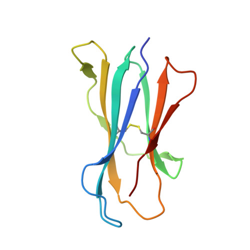
Beta-2 microglobulin99Mus musculusMutation(s): 0 
Gene Names: B2mbeta 2 microglobulinmCG_11606RP23-34E24.5-001
UniProt & NIH Common Fund Data Resources
Find proteins for P01887 (Mus musculus)
Explore P01887 
Go to UniProtKB:  P01887
IMPC:  MGI:88127
Entity Groups  
Sequence Clusters30% Identity50% Identity70% Identity90% Identity95% Identity100% Identity
UniProt GroupP01887
Sequence Annotations
Expand
  • Reference Sequence
Oligosaccharides

Help

Entity ID: 3
MoleculeChains Length2D Diagram Glycosylation3D Interactions
2-acetamido-2-deoxy-beta-D-glucopyranose-(1-4)-2-acetamido-2-deoxy-beta-D-glucopyranose
C
2N-Glycosylation
Glycosylation Resources
GlyTouCan:  G42666HT
GlyCosmos:  G42666HT
GlyGen:  G42666HT
Small Molecules
Ligands 3 Unique
IDChains Name / Formula / InChI Key2D Diagram3D Interactions
FEE
Query on FEE

Download Ideal Coordinates CCD File 
F [auth A]N-{(1S,2S,3R)-1-[(alpha-D-galactopyranosyloxy)methyl]-2,3-dihydroxyoctyl}tetracosanamide
C39 H77 N O9
MPKIDHIOYNMFES-CLTBVUQJSA-N
PLM
Query on PLM

Download Ideal Coordinates CCD File 
G [auth A]PALMITIC ACID
C16 H32 O2
IPCSVZSSVZVIGE-UHFFFAOYSA-N
NAG
Query on NAG

Download Ideal Coordinates CCD File 
D [auth A],
E [auth A]
2-acetamido-2-deoxy-beta-D-glucopyranose
C8 H15 N O6
OVRNDRQMDRJTHS-FMDGEEDCSA-N
Binding Affinity Annotations 
IDSourceBinding Affinity
FEE PDBBind:  3G08 Kd: 6.84 (nM) from 1 assay(s)
Experimental Data & Validation

Experimental Data

  • Method: X-RAY DIFFRACTION
  • Resolution: 1.60 Å
  • R-Value Free: 0.226 
  • R-Value Work: 0.195 
  • R-Value Observed: 0.196 
  • Space Group: P 21 21 21
Unit Cell:
Length ( Å )Angle ( ˚ )
a = 42.24α = 90
b = 107.097β = 90
c = 106.663γ = 90
Software Package:
Software NamePurpose
HKL-2000data collection
MOLREPphasing
REFMACrefinement
HKL-2000data reduction
HKL-2000data scaling

Structure Validation

View Full Validation Report


Ligand Structure Quality Assessment 


Entry History 

Deposition Data

  • Released Date: 2009-12-08 
  • Deposition Author(s): Zajonc, D.M.

Revision History  (Full details and data files)

  • Version 1.0: 2009-12-08
    Type: Initial release
  • Version 1.1: 2011-07-13
    Changes: Non-polymer description, Version format compliance
  • Version 2.0: 2020-07-29
    Type: Remediation
    Reason: Carbohydrate remediation
    Changes: Advisory, Atomic model, Data collection, Database references, Derived calculations, Structure summary
  • Version 2.1: 2023-09-06
    Changes: Data collection, Database references, Refinement description, Structure summary
  • Version 2.2: 2024-10-16
    Changes: Structure summary