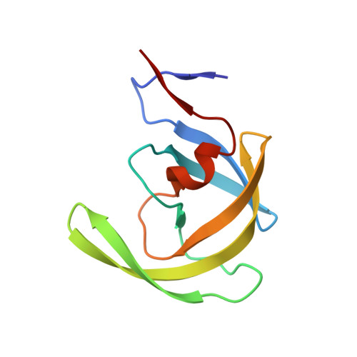
3KFN

HIV Protease (PR) with inhibitor TL-3 and fragment hit 4D9 by soaking


Experimental Data Snapshot

  • Method: X-RAY DIFFRACTION
  • Resolution: 1.77 Å
  • R-Value Free: 0.274 
  • R-Value Work: 0.214 
  • R-Value Observed: 0.217 

Starting Model: experimental
View more details

wwPDB Validation   3D Report Full Report


Ligand Structure Quality Assessment 


This is version 1.4 of the entry. See complete history


Literature

Fragment-based screen against HIV protease.

Perryman, A.L.Zhang, Q.Soutter, H.H.Rosenfeld, R.McRee, D.E.Olson, A.J.Elder, J.E.David Stout, C.

(2010) Chem Biol Drug Des 75: 257-268

  • DOI: https://doi.org/10.1111/j.1747-0285.2009.00943.x
  • Primary Citation of Related Structures:  
    3KF0, 3KFN, 3KFP, 3KFR, 3KFS, 4E43

  • PubMed Abstract: 

    We have employed a fragment-based screen against wild-type (NL4-3) HIV protease (PR) using the Active Sight fragment library and X-ray crystallography. The experiments reveal two new binding sites for small molecules. PR was co-crystallized with fragments, or crystals were soaked in fragment solutions, using five crystal forms, and 378 data sets were collected to 2.3-1.3 A resolution. Fragment binding induces a distinct conformation and specific crystal form of TL-3 inhibited PR during co-crystallization. One fragment, 2-methylcyclohexanol, binds in the 'exo site' adjacent to the Gly(16)Gly(17)Gln(18)loop where the amide of Gly(17)is a specific hydrogen bond donor, and hydrophobic contacts occur with the side chains of Lys(14)and Leu(63). Another fragment, indole-6-carboxylic acid, binds on the 'outside/top of the flap' via hydrophobic contacts with Trp(42), Pro(44), Met(46), and Lys(55), a hydrogen bond with Val(56), and a salt-bridge with Arg(57). 2-acetyl-benzothiophene also binds at this site. This study is the first fragment-based crystallographic screen against HIV PR, and the first time that fragments were screened against an inhibitor-bound drug target to search for compounds that both bind to novel sites and stabilize the inhibited conformation of the target.


  • Organizational Affiliation

    Department of Molecular Biology, The Scripps Research Institute, 10550 N. Torrey Pines Rd., La Jolla, CA 92037, USA.


Macromolecules
Find similar proteins by:  (by identity cutoff)  |  3D Structure
Entity ID: 1
MoleculeChains Sequence LengthOrganismDetailsImage
Protease
A, B
99Human immunodeficiency virus 1Mutation(s): 1 
Gene Names: POL
EC: 3.4.23.16
UniProt
Find proteins for P12499 (Human immunodeficiency virus type 1 group M subtype D (isolate Z2/CDC-Z34))
Explore P12499 
Go to UniProtKB:  P12499
Entity Groups  
Sequence Clusters30% Identity50% Identity70% Identity90% Identity95% Identity100% Identity
UniProt GroupP12499
Sequence Annotations
Expand
  • Reference Sequence
Small Molecules
Ligands 5 Unique
IDChains Name / Formula / InChI Key2D Diagram3D Interactions
3TL
Query on 3TL

Download Ideal Coordinates CCD File 
C [auth A]benzyl [(1S,4S,7S,8R,9R,10S,13S,16S)-7,10-dibenzyl-8,9-dihydroxy-1,16-dimethyl-4,13-bis(1-methylethyl)-2,5,12,15,18-pentaoxo-20-phenyl-19-oxa-3,6,11,14,17-pentaazaicos-1-yl]carbamate
C50 H64 N6 O10
BJJPNOGMLLUCER-KUTQPOQPSA-N
4DX
Query on 4DX

Download Ideal Coordinates CCD File 
D [auth A](1S,2S)-2-methylcyclohexanol
C7 H14 O
NDVWOBYBJYUSMF-BQBZGAKWSA-N
DMS
Query on DMS

Download Ideal Coordinates CCD File 
E [auth A],
F [auth A],
G [auth A],
J [auth B]
DIMETHYL SULFOXIDE
C2 H6 O S
IAZDPXIOMUYVGZ-UHFFFAOYSA-N
BME
Query on BME

Download Ideal Coordinates CCD File 
I [auth B]BETA-MERCAPTOETHANOL
C2 H6 O S
DGVVWUTYPXICAM-UHFFFAOYSA-N
K
Query on K

Download Ideal Coordinates CCD File 
H [auth A],
K [auth B]
POTASSIUM ION
K
NPYPAHLBTDXSSS-UHFFFAOYSA-N
Binding Affinity Annotations 
IDSourceBinding Affinity
3TL BindingDB:  3KFN Ki: 1.5 (nM) from 1 assay(s)
PDBBind:  3KFN IC50: 1.80e+4 (nM) from 1 assay(s)
Biologically Interesting Molecules (External Reference) 1 Unique
Experimental Data & Validation

Experimental Data

  • Method: X-RAY DIFFRACTION
  • Resolution: 1.77 Å
  • R-Value Free: 0.274 
  • R-Value Work: 0.214 
  • R-Value Observed: 0.217 
  • Space Group: P 21 21 2
Unit Cell:
Length ( Å )Angle ( ˚ )
a = 58.95α = 90
b = 85.57β = 90
c = 46.49γ = 90
Software Package:
Software NamePurpose
REFMACrefinement
PDB_EXTRACTdata extraction
Blu-Icedata collection
MOSFLMdata reduction
SCALAdata scaling
MOLREPphasing

Structure Validation

View Full Validation Report


Ligand Structure Quality Assessment 


Entry History 

Deposition Data

  • Released Date: 2010-02-23 
  • Deposition Author(s): Stout, C.D.

Revision History  (Full details and data files)

  • Version 1.0: 2010-02-23
    Type: Initial release
  • Version 1.1: 2011-07-13
    Changes: Atomic model, Database references, Derived calculations, Non-polymer description, Structure summary, Version format compliance
  • Version 1.2: 2013-02-27
    Changes: Other
  • Version 1.3: 2021-10-13
    Changes: Database references, Derived calculations
  • Version 1.4: 2023-09-06
    Changes: Data collection, Refinement description