3L3T

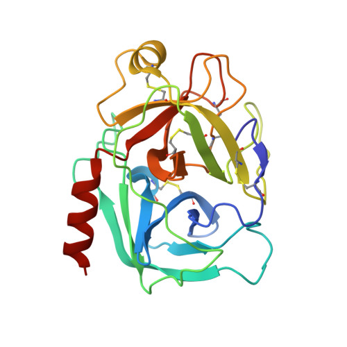
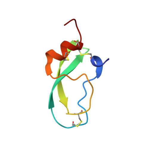
Human mesotrypsin complexed with amyloid precursor protein inhibitor variant (APPIR15K)


Experimental Data Snapshot

  • Method: X-RAY DIFFRACTION
  • Resolution: 2.38 Å
  • R-Value Free: 0.237 
  • R-Value Work: 0.183 

wwPDB Validation   3D Report Full Report


This is version 1.2 of the entry. See complete history


Literature

Determinants of affinity and proteolytic stability in interactions of Kunitz family protease inhibitors with mesotrypsin.

Salameh, M.A.Soares, A.S.Navaneetham, D.Sinha, D.Walsh, P.N.Radisky, E.S.

(2010) J Biol Chem 285: 36884-36896

  • DOI: https://doi.org/10.1074/jbc.M110.171348
  • Primary Citation of Related Structures:  
    3L33, 3L3T

  • PubMed Abstract: 

    An important functional property of protein protease inhibitors is their stability to proteolysis. Mesotrypsin is a human trypsin that has been implicated in the proteolytic inactivation of several protein protease inhibitors. We have found that bovine pancreatic trypsin inhibitor (BPTI), a Kunitz protease inhibitor, inhibits mesotrypsin very weakly and is slowly proteolyzed, whereas, despite close sequence and structural homology, the Kunitz protease inhibitor domain of the amyloid precursor protein (APPI) binds to mesotrypsin 100 times more tightly and is cleaved 300 times more rapidly. To define features responsible for these differences, we have assessed the binding and cleavage by mesotrypsin of APPI and BPTI reciprocally mutated at two nonidentical residues that make direct contact with the enzyme. We find that Arg at P(1) (versus Lys) favors both tighter binding and more rapid cleavage, whereas Met (versus Arg) at P'(2) favors tighter binding but has minimal effect on cleavage. Surprisingly, we find that the APPI scaffold greatly enhances proteolytic cleavage rates, independently of the binding loop. We draw thermodynamic additivity cycles analyzing the interdependence of P(1) and P'(2) substitutions and scaffold differences, finding multiple instances in which the contributions of these features are nonadditive. We also report the crystal structure of the mesotrypsin·APPI complex, in which we find that the binding loop of APPI displays evidence of increased mobility compared with BPTI. Our data suggest that the enhanced vulnerability of APPI to mesotrypsin cleavage may derive from sequence differences in the scaffold that propagate increased flexibility and mobility to the binding loop.


  • Organizational Affiliation

    Department of Cancer Biology, Mayo Clinic Cancer Center, Jacksonville, Florida 32224, USA.


Macromolecules
Find similar proteins by:  (by identity cutoff)  |  3D Structure
Entity ID: 1
MoleculeChains Sequence LengthOrganismDetailsImage
PRSS3 protein
A, B, C, D
224Homo sapiensMutation(s): 1 
Gene Names: PRSS3
EC: 3.4.21.4
UniProt & NIH Common Fund Data Resources
Find proteins for P35030 (Homo sapiens)
Explore P35030 
Go to UniProtKB:  P35030
PHAROS:  P35030
GTEx:  ENSG00000010438 
Entity Groups  
Sequence Clusters30% Identity50% Identity70% Identity90% Identity95% Identity100% Identity
UniProt GroupP35030
Sequence Annotations
Expand
  • Reference Sequence
Find similar proteins by:  (by identity cutoff)  |  3D Structure
Entity ID: 2
MoleculeChains Sequence LengthOrganismDetailsImage
Protein APP
E, F, G, H
57Homo sapiensMutation(s): 1 
Gene Names: APP
Entity Groups  
Sequence Clusters30% Identity50% Identity70% Identity90% Identity95% Identity100% Identity
Sequence Annotations
Expand
  • Reference Sequence
Small Molecules
Ligands 2 Unique
IDChains Name / Formula / InChI Key2D Diagram3D Interactions
FMT
Query on FMT

Download Ideal Coordinates CCD File 
I [auth A]
J [auth A]
K [auth A]
L [auth A]
M [auth A]
I [auth A],
J [auth A],
K [auth A],
L [auth A],
M [auth A],
N [auth A],
O [auth A],
Q [auth B],
R [auth B],
S [auth B],
T [auth B],
U [auth B],
W [auth C]
FORMIC ACID
C H2 O2
BDAGIHXWWSANSR-UHFFFAOYSA-N
CA
Query on CA

Download Ideal Coordinates CCD File 
P [auth A],
V [auth B],
X [auth C],
Y [auth D]
CALCIUM ION
Ca
BHPQYMZQTOCNFJ-UHFFFAOYSA-N
Experimental Data & Validation

Experimental Data

  • Method: X-RAY DIFFRACTION
  • Resolution: 2.38 Å
  • R-Value Free: 0.237 
  • R-Value Work: 0.183 
  • Space Group: P 2 21 21
Unit Cell:
Length ( Å )Angle ( ˚ )
a = 92.793α = 90
b = 130.067β = 90
c = 132.338γ = 90
Software Package:
Software NamePurpose
MOSFLMdata reduction
SCALAdata scaling
PHASERphasing
PHENIXrefinement
PDB_EXTRACTdata extraction

Structure Validation

View Full Validation Report


Entry History 

Deposition Data

Revision History  (Full details and data files)

  • Version 1.0: 2010-09-22
    Type: Initial release
  • Version 1.1: 2011-07-13
    Changes: Version format compliance
  • Version 1.2: 2024-10-30
    Changes: Data collection, Database references, Derived calculations, Structure summary