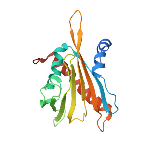
3NJ1

X-ray crystal structure of the PYL2(V114I)-pyrabactin A complex


Experimental Data Snapshot

  • Method: X-RAY DIFFRACTION
  • Resolution: 1.95 Å
  • R-Value Free: 0.217 
  • R-Value Work: 0.195 
  • R-Value Observed: 0.197 

Starting Model: experimental
View more details

wwPDB Validation   3D Report Full Report


Ligand Structure Quality Assessment 


This is version 1.3 of the entry. See complete history


Literature

Structural basis for selective activation of ABA receptors.

Peterson, F.C.Burgie, E.S.Park, S.Y.Jensen, D.R.Weiner, J.J.Bingman, C.A.Chang, C.E.Cutler, S.R.Phillips, G.N.Volkman, B.F.

(2010) Nat Struct Mol Biol 17: 1109-1113

  • DOI: https://doi.org/10.1038/nsmb.1898
  • Primary Citation of Related Structures:  
    3NJ0, 3NJ1, 3NJO

  • PubMed Abstract: 

    Changing environmental conditions and lessening fresh water supplies have sparked intense interest in understanding and manipulating abscisic acid (ABA) signaling, which controls adaptive responses to drought and other abiotic stressors. We recently discovered a selective ABA agonist, pyrabactin, and used it to discover its primary target PYR1, the founding member of the PYR/PYL family of soluble ABA receptors. To understand pyrabactin's selectivity, we have taken a combined structural, chemical and genetic approach. We show that subtle differences between receptor binding pockets control ligand orientation between productive and nonproductive modes. Nonproductive binding occurs without gate closure and prevents receptor activation. Observations in solution show that these orientations are in rapid equilibrium that can be shifted by mutations to control maximal agonist activity. Our results provide a robust framework for the design of new agonists and reveal a new mechanism for agonist selectivity.


  • Organizational Affiliation

    Department of Biochemistry and Center for Eukaryotic Structural Genomics, Medical College of Wisconsin, Milwaukee, Wisconsin, USA.


Macromolecules
Find similar proteins by:  (by identity cutoff)  |  3D Structure
Entity ID: 1
MoleculeChains Sequence LengthOrganismDetailsImage
Abscisic acid receptor PYL2193Arabidopsis thalianaMutation(s): 1 
Gene Names: At2g26040PYL2RCAR14T19L18.15
UniProt
Find proteins for O80992 (Arabidopsis thaliana)
Explore O80992 
Go to UniProtKB:  O80992
Entity Groups  
Sequence Clusters30% Identity50% Identity70% Identity90% Identity95% Identity100% Identity
UniProt GroupO80992
Sequence Annotations
Expand
  • Reference Sequence
Experimental Data & Validation

Experimental Data

  • Method: X-RAY DIFFRACTION
  • Resolution: 1.95 Å
  • R-Value Free: 0.217 
  • R-Value Work: 0.195 
  • R-Value Observed: 0.197 
  • Space Group: P 61 2 2
Unit Cell:
Length ( Å )Angle ( ˚ )
a = 61.754α = 90
b = 61.754β = 90
c = 222.002γ = 120
Software Package:
Software NamePurpose
DENZOdata reduction
SCALEPACKdata scaling
PHASERphasing
PHENIXrefinement
PDB_EXTRACTdata extraction

Structure Validation

View Full Validation Report


Ligand Structure Quality Assessment 


Entry History 

Revision History  (Full details and data files)

  • Version 1.0: 2010-08-18
    Type: Initial release
  • Version 1.1: 2011-07-13
    Changes: Version format compliance
  • Version 1.2: 2017-11-08
    Changes: Refinement description
  • Version 1.3: 2023-09-06
    Changes: Data collection, Database references, Derived calculations, Refinement description