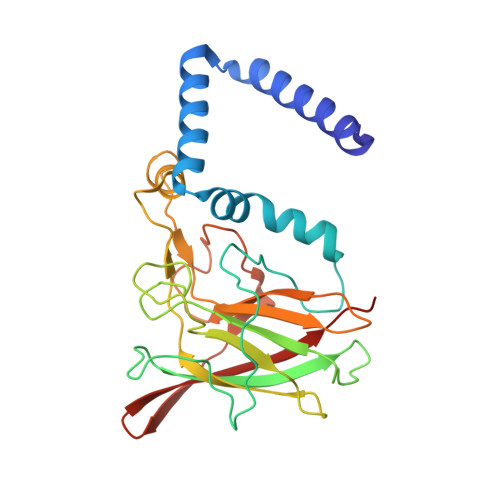
3O5U

Crystal Structure of 4-Chlorocatechol Dioxygenase from Rhodococcus opacus 1CP in complex with protocatechuate


Experimental Data Snapshot

  • Method: X-RAY DIFFRACTION
  • Resolution: 2.35 Å
  • R-Value Free: 0.216 
  • R-Value Work: 0.166 
  • R-Value Observed: 0.169 

Starting Model: experimental
View more details

wwPDB Validation   3D Report Full Report


Ligand Structure Quality Assessment 


This is version 1.1 of the entry. See complete history


Literature

X-ray structures of 4-chlorocatechol 1,2-dioxygenase adducts with substituted catechols: new perspectives in the molecular basis of intradiol ring cleaving dioxygenases specificity.

Ferraroni, M.Kolomytseva, M.Scozzafava, A.Golovleva, L.Briganti, F.

(2013) J Struct Biol 181: 274-282

  • DOI: https://doi.org/10.1016/j.jsb.2012.11.007
  • Primary Citation of Related Structures:  
    3O32, 3O5U, 3O6J, 3O6R

  • PubMed Abstract: 

    The crystallographic structures of 4-chlorocatechol 1,2-dioxygenase (4-CCD) complexes with 3,5-dichlorocatechol, protocatechuate (3,4-dihydroxybenzoate), hydroxyquinol (benzen-1,2,4-triol) and pyrogallol (benzen-1,2,3-triol), which act as substrates or inhibitors of the enzyme, have been determined and analyzed. 4-CCD from the Gram-positive bacterium Rhodococcus opacus 1CP is a Fe(III) ion containing enzyme specialized in the aerobic biodegradation of chlorocatechols. The structures of the 4-CCD complexes show that the catechols bind the catalytic iron ion in a bidentate mode displacing Tyr169 and the benzoate ion (found in the native enzyme structure) from the metal coordination sphere, as found in other adducts of intradiol dioxygenases with substrates. The analysis of the present structures allowed to identify the residues selectively involved in recognition of the diverse substrates. Furthermore the structural comparison with the corresponding complexes of catechol 1,2-dioxygenase from the same Rhodococcus strain (Rho-1,2-CTD) highlights significant differences in the binding of the tested catechols to the active site of the enzyme, particularly in the orientation of the aromatic ring substituents. As an example the 3-substituted catechols are bound with the substituent oriented towards the external part of the 4-CCD active site cavity, whereas in the Rho-1,2-CTD complexes the 3-substituents were placed in the internal position. The present crystallographic study shed light on the mechanism that allows substrate recognition inside this class of high specific enzymes involved in the biodegradation of recalcitrant pollutants.


  • Organizational Affiliation

    Dipartimento di Chimica "Ugo Schiff", Università di Firenze, Via della Lastruccia 3, I-50019 Sesto Fiorentino (FI), Italy. marta.ferraroni@unifi.it


Macromolecules
Find similar proteins by:  (by identity cutoff)  |  3D Structure
Entity ID: 1
MoleculeChains Sequence LengthOrganismDetailsImage
Chlorocatechol 1,2-dioxygenase
A, B
257Rhodococcus opacusMutation(s): 0 
EC: 1.13.11
UniProt
Find proteins for O67987 (Rhodococcus opacus)
Explore O67987 
Go to UniProtKB:  O67987
Entity Groups  
Sequence Clusters30% Identity50% Identity70% Identity90% Identity95% Identity100% Identity
UniProt GroupO67987
Sequence Annotations
Expand
  • Reference Sequence
Small Molecules
Ligands 5 Unique
IDChains Name / Formula / InChI Key2D Diagram3D Interactions
MYY
Query on MYY

Download Ideal Coordinates CCD File 
D [auth A],
E [auth A]
(2R)-3-(PHOSPHONOOXY)-2-(TETRADECANOYLOXY)PROPYL PALMITATE
C33 H65 O8 P
GLOXZZHEZYKXNV-WJOKGBTCSA-N
DHB
Query on DHB

Download Ideal Coordinates CCD File 
F [auth A],
M [auth B]
3,4-DIHYDROXYBENZOIC ACID
C7 H6 O4
YQUVCSBJEUQKSH-UHFFFAOYSA-N
GOL
Query on GOL

Download Ideal Coordinates CCD File 
G [auth A],
H [auth A],
N [auth B]
GLYCEROL
C3 H8 O3
PEDCQBHIVMGVHV-UHFFFAOYSA-N
FE
Query on FE

Download Ideal Coordinates CCD File 
C [auth A],
L [auth B]
FE (III) ION
Fe
VTLYFUHAOXGGBS-UHFFFAOYSA-N
CL
Query on CL

Download Ideal Coordinates CCD File 
I [auth A],
J [auth A],
K [auth A],
O [auth B]
CHLORIDE ION
Cl
VEXZGXHMUGYJMC-UHFFFAOYSA-M
Experimental Data & Validation

Experimental Data

  • Method: X-RAY DIFFRACTION
  • Resolution: 2.35 Å
  • R-Value Free: 0.216 
  • R-Value Work: 0.166 
  • R-Value Observed: 0.169 
  • Space Group: P 63
Unit Cell:
Length ( Å )Angle ( ˚ )
a = 87.31α = 90
b = 87.31β = 90
c = 185.921γ = 120
Software Package:
Software NamePurpose
MOSFLMdata reduction
SCALAdata scaling
REFMACrefinement
PDB_EXTRACTdata extraction
MAR345dtbdata collection
MOLREPphasing

Structure Validation

View Full Validation Report


Ligand Structure Quality Assessment 


Entry History 

Deposition Data

Revision History  (Full details and data files)

  • Version 1.0: 2011-08-17
    Type: Initial release
  • Version 1.1: 2023-09-06
    Changes: Data collection, Database references, Derived calculations, Refinement description