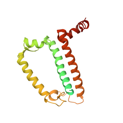
3PL9 | pdb_00003pl9

Crystal structure of spinach minor light-harvesting complex CP29 at 2.80 angstrom resolution


Experimental Data Snapshot

  • Method: X-RAY DIFFRACTION
  • Resolution: 2.80 Å
  • R-Value Free: 
    0.293 (Depositor), 0.280 (DCC) 
  • R-Value Work: 
    0.282 (Depositor), 0.280 (DCC) 
  • R-Value Observed: 
    0.282 (Depositor) 

wwPDB Validation   3D Report Full Report


Ligand Structure Quality Assessment 

Created with Raphaël 2.3.0Worse 01 BetterLigand structure goodness of fit to experimental dataBest fitted CHLClick on this verticalbar to view detailsBest fitted CLAClick on this verticalbar to view detailsBest fitted NEXClick on this verticalbar to view detailsBest fitted XATClick on this verticalbar to view detailsBest fitted LUTClick on this verticalbar to view detailsBest fitted HTGClick on this verticalbar to view detailsBest fitted G3PClick on this verticalbar to view details

This is version 3.0 of the entry. See complete history


Literature

Structural insights into energy regulation of light-harvesting complex CP29 from spinach.

Pan, X.Li, M.Wan, T.Wang, L.Jia, C.Hou, Z.Zhao, X.Zhang, J.Chang, W.

(2011) Nat Struct Mol Biol 18: 309-315

  • DOI: https://doi.org/10.1038/nsmb.2008
  • Primary Citation of Related Structures:  
    3PL9

  • PubMed Abstract: 

    CP29, one of the minor light-harvesting complexes of higher-plant photosystem II, absorbs and transfers solar energy for photosynthesis and also has important roles in photoprotection. We have solved the crystal structure of spinach CP29 at 2.80-Å resolution. Each CP29 monomer contains 13 chlorophyll and 3 carotenoid molecules, which differs considerably from the major light-harvesting complex LHCII and the previously proposed CP29 model. The 13 chlorophyll-binding sites are assigned as eight chlorophyll a sites, four chlorophyll b and one putative mixed site occupied by both chlorophylls a and b. Based on the present X-ray structure, an integrated pigment network in CP29 is constructed. Two special clusters of pigment molecules, namely a615-a611-a612-Lut and Vio(Zea)-a603-a609, have been identified and might function as potential energy-quenching centers and as the exit or entrance in energy-transfer pathways.


  • Organizational Affiliation

    National Laboratory of Biomacromolecules, Institute of Biophysics, Chinese Academy of Sciences, Beijing, China.


Macromolecules
Find similar proteins by:  (by identity cutoff)  |  3D Structure
Entity ID: 1
MoleculeChains Sequence LengthOrganismDetailsImage
Chlorophyll A-B binding protein243Spinacia oleraceaMutation(s): 0 
UniProt
Find proteins for F2Z293 (Spinacia oleracea)
Explore F2Z293 
Go to UniProtKB:  F2Z293
Entity Groups  
Sequence Clusters30% Identity50% Identity70% Identity90% Identity95% Identity100% Identity
UniProt GroupF2Z293
Sequence Annotations
Expand
  • Reference Sequence
Small Molecules
Ligands 7 Unique
IDChains Name / Formula / InChI Key2D Diagram3D Interactions
CHL
Query on CHL

Download Ideal Coordinates CCD File 
E [auth A],
F [auth A],
G [auth A],
M [auth A]
CHLOROPHYLL B
C55 H70 Mg N4 O6
MWVCRINOIIOUAU-UYSPMESUSA-M
CLA
Query on CLA

Download Ideal Coordinates CCD File 
B [auth A]
C [auth A]
D [auth A]
H [auth A]
I [auth A]
CHLOROPHYLL A
C55 H72 Mg N4 O5
ATNHDLDRLWWWCB-AENOIHSZSA-M
NEX
Query on NEX

Download Ideal Coordinates CCD File 
Q [auth A](1R,3R)-6-{(3E,5E,7E,9E,11E,13E,15E,17E)-18-[(1S,4R,6R)-4-HYDROXY-2,2,6-TRIMETHYL-7-OXABICYCLO[4.1.0]HEPT-1-YL]-3,7,12,16-TETRAMETHYLOCTADECA-1,3,5,7,9,11,13,15,17-NONAENYLIDENE}-1,5,5-TRIMETHYLCYCLOHEXANE-1,3-DIOL
C40 H56 O4
PGYAYSRVSAJXTE-OQASCVKESA-N
XAT
Query on XAT

Download Ideal Coordinates CCD File 
P [auth A](3S,5R,6S,3'S,5'R,6'S)-5,6,5',6'-DIEPOXY-5,6,5',6'- TETRAHYDRO-BETA,BETA-CAROTENE-3,3'-DIOL
C40 H56 O4
SZCBXWMUOPQSOX-WVJDLNGLSA-N
LUT
Query on LUT

Download Ideal Coordinates CCD File 
O [auth A](3R,3'R,6S)-4,5-DIDEHYDRO-5,6-DIHYDRO-BETA,BETA-CAROTENE-3,3'-DIOL
C40 H56 O2
KBPHJBAIARWVSC-NSIPBSJQSA-N
HTG
Query on HTG

Download Ideal Coordinates CCD File 
S [auth A],
T [auth A]
heptyl 1-thio-beta-D-glucopyranoside
C13 H26 O5 S
HPEGNLMTTNTJSP-LBELIVKGSA-N
G3P
Query on G3P

Download Ideal Coordinates CCD File 
R [auth A]SN-GLYCEROL-3-PHOSPHATE
C3 H9 O6 P
AWUCVROLDVIAJX-GSVOUGTGSA-N
Experimental Data & Validation

Experimental Data

  • Method: X-RAY DIFFRACTION
  • Resolution: 2.80 Å
  • R-Value Free:  0.293 (Depositor), 0.280 (DCC) 
  • R-Value Work:  0.282 (Depositor), 0.280 (DCC) 
  • R-Value Observed: 0.282 (Depositor) 
Space Group: P 65 2 2
Unit Cell:
Length ( Å )Angle ( ˚ )
a = 68.793α = 90
b = 68.793β = 90
c = 425.778γ = 120
Software Package:
Software NamePurpose
HKL-2000data collection
SOLVEphasing
CNSrefinement
HKL-2000data reduction
HKL-2000data scaling

Structure Validation

View Full Validation Report


Ligand Structure Quality Assessment 

Created with Raphaël 2.3.0Worse 01 BetterLigand structure goodness of fit to experimental dataBest fitted CHLClick on this verticalbar to view detailsBest fitted CLAClick on this verticalbar to view detailsBest fitted NEXClick on this verticalbar to view detailsBest fitted XATClick on this verticalbar to view detailsBest fitted LUTClick on this verticalbar to view detailsBest fitted HTGClick on this verticalbar to view detailsBest fitted G3PClick on this verticalbar to view details

Entry History 

Deposition Data

Revision History  (Full details and data files)

  • Version 1.0: 2011-02-09
    Type: Initial release
  • Version 1.1: 2011-07-13
    Changes: Version format compliance
  • Version 2.0: 2019-05-01
    Changes: Advisory, Atomic model, Data collection, Derived calculations
  • Version 2.1: 2019-12-25
    Changes: Database references
  • Version 2.2: 2020-07-29
    Type: Remediation
    Reason: Carbohydrate remediation
    Changes: Derived calculations, Structure summary
  • Version 3.0: 2024-03-20
    Changes: Data collection, Database references, Non-polymer description, Structure summary