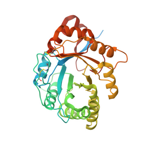
3RLH

Crystal structure of a class II phospholipase D from Loxosceles intermedia venom


Experimental Data Snapshot

  • Method: X-RAY DIFFRACTION
  • Resolution: 1.72 Å
  • R-Value Free: 0.214 
  • R-Value Work: 0.172 
  • R-Value Observed: 0.174 

Starting Model: experimental
View more details

wwPDB Validation   3D Report Full Report


This is version 1.3 of the entry. See complete history


Literature

Structure of a novel class II phospholipase D: Catalytic cleft is modified by a disulphide bridge.

de Giuseppe, P.O.Ullah, A.Silva, D.T.Gremski, L.H.Wille, A.C.Chaves Moreira, D.Ribeiro, A.S.Chaim, O.M.Murakami, M.T.Veiga, S.S.Arni, R.K.

(2011) Biochem Biophys Res Commun 409: 622-627

  • DOI: https://doi.org/10.1016/j.bbrc.2011.05.053
  • Primary Citation of Related Structures:  
    3RLH

  • PubMed Abstract: 

    Phospholipases D (PLDs) are principally responsible for the local and systemic effects of Loxosceles envenomation including dermonecrosis and hemolysis. Despite their clinical relevance in loxoscelism, to date, only the SMase I from Loxosceles laeta, a class I member, has been structurally characterized. The crystal structure of a class II member from Loxosceles intermedia venom has been determined at 1.7Å resolution. Structural comparison to the class I member showed that the presence of an additional disulphide bridge which links the catalytic loop to the flexible loop significantly changes the volume and shape of the catalytic cleft. An examination of the crystal structures of PLD homologues in the presence of low molecular weight compounds at their active sites suggests the existence of a ligand-dependent rotamer conformation of the highly conserved residue Trp230 (equivalent to Trp192 in the glycerophosphodiester phosphodiesterase from Thermus thermophofilus, PDB code: 1VD6) indicating its role in substrate binding in both enzymes. Sequence and structural analyses suggest that the reduced sphingomyelinase activity observed in some class IIb PLDs is probably due to point mutations which lead to a different substrate preference.


  • Organizational Affiliation

    Laboratório Nacional de Biociências, Centro Nacional de Pesquisa em Energia e Materiais, Campinas, 13083-970 SP, Brazil.


Macromolecules
Find similar proteins by:  (by identity cutoff)  |  3D Structure
Entity ID: 1
MoleculeChains Sequence LengthOrganismDetailsImage
Sphingomyelin phosphodiesterase D LiSicTox-alphaIA1a329Loxosceles intermediaMutation(s): 1 
EC: 3.1.4.41 (PDB Primary Data), 4.6.1 (UniProt)
Membrane Entity: Yes 
UniProt
Find proteins for P0CE80 (Loxosceles intermedia)
Explore P0CE80 
Go to UniProtKB:  P0CE80
Entity Groups  
Sequence Clusters30% Identity50% Identity70% Identity90% Identity95% Identity100% Identity
UniProt GroupP0CE80
Sequence Annotations
Expand
  • Reference Sequence
Small Molecules
Ligands 4 Unique
IDChains Name / Formula / InChI Key2D Diagram3D Interactions
PGE
Query on PGE

Download Ideal Coordinates CCD File 
C [auth A]TRIETHYLENE GLYCOL
C6 H14 O4
ZIBGPFATKBEMQZ-UHFFFAOYSA-N
PEG
Query on PEG

Download Ideal Coordinates CCD File 
D [auth A],
E [auth A],
F [auth A],
G [auth A]
DI(HYDROXYETHYL)ETHER
C4 H10 O3
MTHSVFCYNBDYFN-UHFFFAOYSA-N
EDO
Query on EDO

Download Ideal Coordinates CCD File 
H [auth A],
I [auth A]
1,2-ETHANEDIOL
C2 H6 O2
LYCAIKOWRPUZTN-UHFFFAOYSA-N
MG
Query on MG

Download Ideal Coordinates CCD File 
B [auth A]MAGNESIUM ION
Mg
JLVVSXFLKOJNIY-UHFFFAOYSA-N
Experimental Data & Validation

Experimental Data

  • Method: X-RAY DIFFRACTION
  • Resolution: 1.72 Å
  • R-Value Free: 0.214 
  • R-Value Work: 0.172 
  • R-Value Observed: 0.174 
  • Space Group: P 1 21 1
Unit Cell:
Length ( Å )Angle ( ˚ )
a = 49.81α = 90
b = 49.3β = 105.83
c = 56.3γ = 90
Software Package:
Software NamePurpose
MOSFLMdata reduction
SCALEPACKdata scaling
MOLREPphasing
REFMACrefinement
PDB_EXTRACTdata extraction
MAR345dtbdata collection
SCALAdata scaling

Structure Validation

View Full Validation Report


Entry History 

Deposition Data

Revision History  (Full details and data files)

  • Version 1.0: 2011-06-29
    Type: Initial release
  • Version 1.1: 2011-07-13
    Changes: Version format compliance
  • Version 1.2: 2023-09-13
    Changes: Data collection, Database references, Derived calculations, Refinement description
  • Version 1.3: 2024-11-06
    Changes: Structure summary