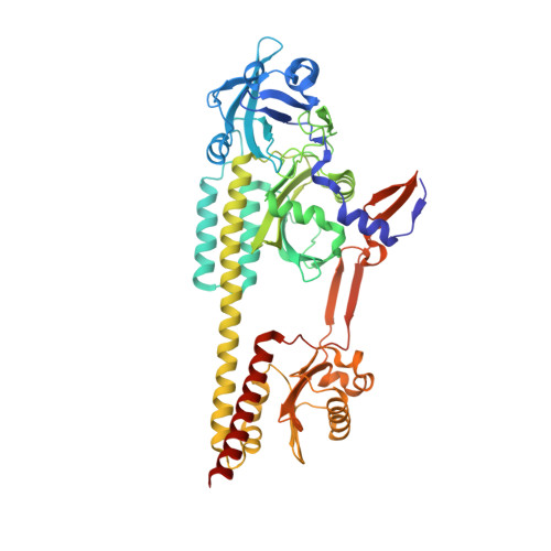
3ZQ5

Structure of the Y263F mutant of the cyanobacterial phytochrome Cph1


Experimental Data Snapshot

  • Method: X-RAY DIFFRACTION
  • Resolution: 1.95 Å
  • R-Value Free: 0.218 
  • R-Value Work: 0.174 
  • R-Value Observed: 0.176 

Starting Model: experimental
View more details

wwPDB Validation   3D Report Full Report


Ligand Structure Quality Assessment 


This is version 1.3 of the entry. See complete history


Literature

Spectroscopy and a High-Resolution Crystal Structure of Tyr263 Mutants of Cyanobacterial Phytochrome Cph1.

Mailliet, J.Psakis, G.Feilke, K.Sineshchekov, V.Essen, L.-O.Hughes, J.

(2011) J Mol Biol 413: 115

  • DOI: https://doi.org/10.1016/j.jmb.2011.08.023
  • Primary Citation of Related Structures:  
    3ZQ5

  • PubMed Abstract: 

    Phytochromes are biliprotein photoreceptors that can be photoswitched between red-light-absorbing state (Pr) and far-red-light-absorbing state (Pfr). Although three-dimensional structures of both states have been reported, the photoconversion and intramolecular signaling mechanisms are still unclear. Here, we report UV-Vis absorbance, fluorescence and CD spectroscopy along with various photochemical parameters of the wild type and Y263F, Y263H and Y263S mutants of the Cph1 photosensory module, as well as a 2.0-Å-resolution crystal structure of the Y263F mutant in its Pr ground state. Although Y263 is conserved, we show that the aromatic character but not the hydroxyl group of Y263 is important for Pfr formation. The crystal structure of the Y263F mutant (Protein Data Bank ID: 3ZQ5) reaffirms the ZZZssa chromophore configuration and provides a detailed picture of its binding pocket, particularly conformational heterogeneity around the chromophore. Comparison with other phytochrome structures reveals differences in the relative position of the PHY (phytochrome specific) domain and the interaction of the tongue with the extreme N-terminus. Our data support the notion that native phytochromes in their Pr state are structurally heterogeneous.


  • Organizational Affiliation

    Institute for Plant Physiology, Justus Liebig University Giessen, Senckenbergstrasse 3, D35390 Giessen, Germany.


Macromolecules
Find similar proteins by:  (by identity cutoff)  |  3D Structure
Entity ID: 1
MoleculeChains Sequence LengthOrganismDetailsImage
PHYTOCHROME-LIKE PROTEIN CPH1520Synechocystis sp. PCC 6803Mutation(s): 1 
EC: 2.7.13.3
UniProt
Find proteins for Q55168 (Synechocystis sp. (strain ATCC 27184 / PCC 6803 / Kazusa))
Explore Q55168 
Go to UniProtKB:  Q55168
Entity Groups  
Sequence Clusters30% Identity50% Identity70% Identity90% Identity95% Identity100% Identity
UniProt GroupQ55168
Sequence Annotations
Expand
  • Reference Sequence
Small Molecules
Ligands 5 Unique
IDChains Name / Formula / InChI Key2D Diagram3D Interactions
CYC
Query on CYC

Download Ideal Coordinates CCD File 
B [auth A]PHYCOCYANOBILIN
C33 H40 N4 O6
VXTXPYZGDQPMHK-GMXXPEQVSA-N
PO4
Query on PO4

Download Ideal Coordinates CCD File 
D [auth A]PHOSPHATE ION
O4 P
NBIIXXVUZAFLBC-UHFFFAOYSA-K
GOL
Query on GOL

Download Ideal Coordinates CCD File 
I [auth A],
J [auth A]
GLYCEROL
C3 H8 O3
PEDCQBHIVMGVHV-UHFFFAOYSA-N
ACT
Query on ACT

Download Ideal Coordinates CCD File 
E [auth A],
F [auth A],
G [auth A],
H [auth A]
ACETATE ION
C2 H3 O2
QTBSBXVTEAMEQO-UHFFFAOYSA-M
NA
Query on NA

Download Ideal Coordinates CCD File 
C [auth A],
K [auth A]
SODIUM ION
Na
FKNQFGJONOIPTF-UHFFFAOYSA-N
Experimental Data & Validation

Experimental Data

  • Method: X-RAY DIFFRACTION
  • Resolution: 1.95 Å
  • R-Value Free: 0.218 
  • R-Value Work: 0.174 
  • R-Value Observed: 0.176 
  • Space Group: C 1 2 1
Unit Cell:
Length ( Å )Angle ( ˚ )
a = 107.57α = 90
b = 95.15β = 99.67
c = 73.58γ = 90
Software Package:
Software NamePurpose
REFMACrefinement
XDSdata reduction
XSCALEdata scaling
PHASERphasing

Structure Validation

View Full Validation Report


Ligand Structure Quality Assessment 


Entry History 

Deposition Data

Revision History  (Full details and data files)

  • Version 1.0: 2011-11-30
    Type: Initial release
  • Version 1.1: 2018-02-07
    Changes: Source and taxonomy
  • Version 1.2: 2023-12-20
    Changes: Data collection, Database references, Derived calculations, Other, Refinement description
  • Version 1.3: 2024-10-23
    Changes: Structure summary