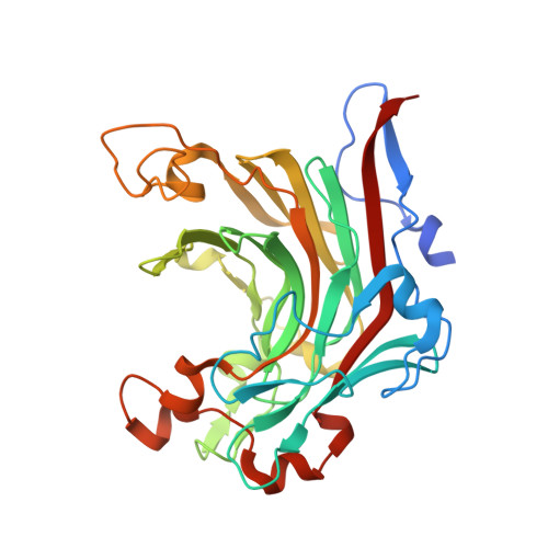
4ATF

Crystal structure of inactivated mutant beta-agarase B in complex with agaro-octaose


Experimental Data Snapshot

  • Method: X-RAY DIFFRACTION
  • Resolution: 1.90 Å
  • R-Value Free: 0.190 
  • R-Value Work: 0.134 
  • R-Value Observed: 0.137 

Starting Model: experimental
View more details

wwPDB Validation   3D Report Full Report


This is version 2.1 of the entry. See complete history


Literature

Biochemical and Structural Characterization of the Complex Agarolytic Enzyme System from the Marine Bacterium Zobellia Galactanivorans.

Hehemann, J.H.Correc, G.Thomas, F.Bernard, T.Barbeyron, T.Jam, M.Helbert, W.Michel, G.Czjzek, M.

(2012) J Biol Chem 287: 30571

  • DOI: https://doi.org/10.1074/jbc.M112.377184
  • Primary Citation of Related Structures:  
    4ASM, 4ATE, 4ATF

  • PubMed Abstract: 

    Zobellia galactanivorans is an emerging model bacterium for the bioconversion of algal biomass. Notably, this marine Bacteroidetes possesses a complex agarolytic system comprising four β-agarases and five β-porphyranases, all belonging to the glycoside hydrolase family 16. Although β-agarases are specific for the neutral agarobiose moieties, the recently discovered β-porphyranases degrade the sulfated polymers found in various quantities in natural agars. Here, we report the biochemical and structural comparison of five β-porphyranases and β-agarases from Z. galactanivorans. The respective degradation patterns of two β-porphyranases and three β-agarases are analyzed by their action on defined hybrid oligosaccharides. In light of the high resolution crystal structures, the biochemical results allowed a detailed mapping of substrate specificities along the active site groove of the enzymes. Although PorA displays a strict requirement for C6-sulfate in the -2- and +1-binding subsites, PorB tolerates the presence of 3-6-anhydro-l-galactose in subsite -2. Both enzymes do not accept methylation of the galactose unit in the -1 subsite. The β-agarase AgaD requires at least four consecutive agarose units (DP8) and is highly intolerant to modifications, whereas for AgaB oligosaccharides containing C6-sulfate groups at the -4, +1, and +3 positions are still degraded. Together with a transcriptional analysis of the expression of these enzymes, the structural and biochemical results allow proposition of a model scheme for the agarolytic system of Z. galactanivorans.


  • Organizational Affiliation

    Université Pierre et Marie Curie, Végétaux Marins et Biomolécules UMR 7139, Station Biologique de Roscoff, F 29682 Roscoff, France.


Macromolecules
Find similar proteins by:  (by identity cutoff)  |  3D Structure
Entity ID: 1
MoleculeChains Sequence LengthOrganismDetailsImage
BETA-AGARASE B
A, B, C, D
308Zobellia galactanivoransMutation(s): 1 
EC: 3.2.1.81
UniProt
Find proteins for Q9RGX8 (Zobellia galactanivorans (strain DSM 12802 / CCUG 47099 / CIP 106680 / NCIMB 13871 / Dsij))
Explore Q9RGX8 
Go to UniProtKB:  Q9RGX8
Entity Groups  
Sequence Clusters30% Identity50% Identity70% Identity90% Identity95% Identity100% Identity
UniProt GroupQ9RGX8
Sequence Annotations
Expand
  • Reference Sequence
Oligosaccharides

Help

Entity ID: 2
MoleculeChains Length2D Diagram Glycosylation3D Interactions
3,6-anhydro-alpha-L-galactopyranose-(1-3)-beta-D-galactopyranose-(1-4)-3,6-anhydro-alpha-L-galactopyranose-(1-3)-beta-D-galactopyranose-(1-4)-3,6-anhydro-alpha-L-galactopyranose-(1-3)-beta-D-galactopyranose-(1-4)-3,6-anhydro-alpha-L-galactopyranose-(1-3)-beta-D-galactopyranose
E, F, G, H
8N/A
Glycosylation Resources
GlyTouCan:  G63389KU
GlyCosmos:  G63389KU
Experimental Data & Validation

Experimental Data

  • Method: X-RAY DIFFRACTION
  • Resolution: 1.90 Å
  • R-Value Free: 0.190 
  • R-Value Work: 0.134 
  • R-Value Observed: 0.137 
  • Space Group: P 1 21 1
Unit Cell:
Length ( Å )Angle ( ˚ )
a = 73.187α = 90
b = 106.234β = 93.22
c = 96.983γ = 90
Software Package:
Software NamePurpose
REFMACrefinement
MOSFLMdata reduction
SCALAdata scaling
AMoREphasing

Structure Validation

View Full Validation Report


Entry History 

Deposition Data

Revision History  (Full details and data files)

  • Version 1.0: 2012-07-25
    Type: Initial release
  • Version 1.1: 2012-09-12
    Changes: Database references, Structure summary
  • Version 1.2: 2017-08-09
    Changes: Data collection
  • Version 2.0: 2020-07-29
    Type: Remediation
    Reason: Carbohydrate remediation
    Changes: Advisory, Atomic model, Data collection, Derived calculations, Other, Structure summary
  • Version 2.1: 2023-12-20
    Changes: Data collection, Database references, Refinement description, Structure summary