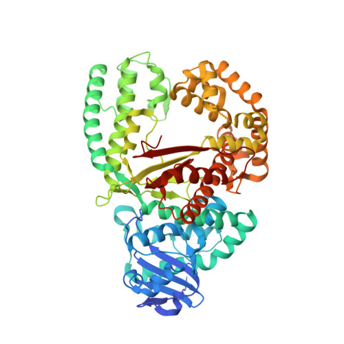
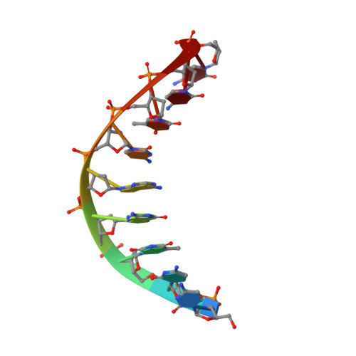
4DQP

Ternary complex of Bacillus DNA Polymerase I Large Fragment, DNA duplex, and ddCTP (paired with dG of template)


Experimental Data Snapshot

  • Method: X-RAY DIFFRACTION
  • Resolution: 1.74 Å
  • R-Value Free: 0.195 
  • R-Value Work: 0.167 
  • R-Value Observed: 0.168 

Starting Model: experimental
View more details

wwPDB Validation   3D Report Full Report


Ligand Structure Quality Assessment 


This is version 1.3 of the entry. See complete history


Literature

Structural factors that determine selectivity of a high fidelity DNA polymerase for deoxy-, dideoxy-, and ribonucleotides.

Wang, W.Wu, E.Y.Hellinga, H.W.Beese, L.S.

(2012) J Biol Chem 287: 28215-28226

  • DOI: https://doi.org/10.1074/jbc.M112.366609
  • Primary Citation of Related Structures:  
    4DQI, 4DQP, 4DQQ, 4DQR, 4DQS, 4DS4, 4DS5, 4DSE, 4DSF, 4E0D

  • PubMed Abstract: 

    In addition to discriminating against base pair mismatches, DNA polymerases exhibit a high degree of selectivity for deoxyribonucleotides over ribo- or dideoxynucleotides. It has been proposed that a single active site residue (steric gate) blocks productive binding of nucleotides containing 2'-hydroxyls. Although this steric gate plays a role in sugar moiety discrimination, its interactions do not account fully for the observed behavior of mutants. Here we present 10 high resolution crystal structures and enzyme kinetic analyses of Bacillus DNA polymerase I large fragment variants complexed with deoxy-, ribo-, and dideoxynucleotides and a DNA substrate. Taken together, these data present a more nuanced and general mechanism for nucleotide discrimination in which ensembles of intermediate conformations in the active site trap non-cognate substrates. It is known that the active site O-helix transitions from an open state in the absence of nucleotide substrates to a ternary complex closed state in which the reactive groups are aligned for catalysis. Substrate misalignment in the closed state plays a fundamental part in preventing non-cognate nucleotide misincorpation. The structures presented here show that additional O-helix conformations intermediate between the open and closed state extremes create an ensemble of binding sites that trap and misalign non-cognate nucleotides. Water-mediated interactions, absent in the fully closed state, play an important role in formation of these binding sites and can be remodeled to accommodate different non-cognate substrates. This mechanism may extend also to base pair discrimination.


  • Organizational Affiliation

    Department of Biochemistry, Duke University Medical Center, Durham, North Carolina 27710, USA.


Macromolecules

Find similar proteins by:  (by identity cutoff)  |  3D Structure
Entity ID: 1
MoleculeChains Sequence LengthOrganismDetailsImage
DNA polymeraseA,
B [auth D]
592Geobacillus kaustophilus HTA426Mutation(s): 1 
Gene Names: polAGK2730
EC: 2.7.7.7
UniProt
Find proteins for Q5KWC1 (Geobacillus kaustophilus (strain HTA426))
Explore Q5KWC1 
Go to UniProtKB:  Q5KWC1
Entity Groups  
Sequence Clusters30% Identity50% Identity70% Identity90% Identity95% Identity100% Identity
UniProt GroupQ5KWC1
Sequence Annotations
Expand
  • Reference Sequence

Find similar nucleic acids by:  Sequence   |   3D Structure  

Entity ID: 2
MoleculeChains LengthOrganismImage
DNA (5'-D(*CP*CP*TP*GP*AP*CP*TP*CP*(DOC))-3')C [auth B],
E
9N/A
Sequence Annotations
Expand
  • Reference Sequence

Find similar nucleic acids by:  Sequence   |   3D Structure  

Entity ID: 3
MoleculeChains LengthOrganismImage
DNA (5'-D(*C*AP*TP*GP*GP*GP*AP*GP*TP*CP*AP*GP*G)-3')D [auth C],
F
13N/A
Sequence Annotations
Expand
  • Reference Sequence
Small Molecules
Ligands 3 Unique
IDChains Name / Formula / InChI Key2D Diagram3D Interactions
DCT
Query on DCT

Download Ideal Coordinates CCD File 
G [auth A],
H [auth A],
J [auth D]
2',3'-DIDEOXYCYTIDINE 5'-TRIPHOSPHATE
C9 H16 N3 O12 P3
ARLKCWCREKRROD-POYBYMJQSA-N
SO4
Query on SO4

Download Ideal Coordinates CCD File 
L [auth D],
M [auth D]
SULFATE ION
O4 S
QAOWNCQODCNURD-UHFFFAOYSA-L
MG
Query on MG

Download Ideal Coordinates CCD File 
I [auth A],
K [auth D]
MAGNESIUM ION
Mg
JLVVSXFLKOJNIY-UHFFFAOYSA-N
Experimental Data & Validation

Experimental Data

  • Method: X-RAY DIFFRACTION
  • Resolution: 1.74 Å
  • R-Value Free: 0.195 
  • R-Value Work: 0.167 
  • R-Value Observed: 0.168 
  • Space Group: P 21 21 21
Unit Cell:
Length ( Å )Angle ( ˚ )
a = 94.22α = 90
b = 109.34β = 90
c = 149.93γ = 90
Software Package:
Software NamePurpose
XSCALEdata scaling
PHENIXrefinement
PDB_EXTRACTdata extraction
XDSdata reduction

Structure Validation

View Full Validation Report


Ligand Structure Quality Assessment 


Entry History 

Deposition Data

Revision History  (Full details and data files)

  • Version 1.0: 2012-06-06
    Type: Initial release
  • Version 1.1: 2012-06-13
    Changes: Database references
  • Version 1.2: 2012-11-21
    Changes: Database references
  • Version 1.3: 2023-09-13
    Changes: Data collection, Database references, Derived calculations, Refinement description