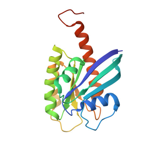
4DST

Small-molecule ligands bind to a distinct pocket in Ras and inhibit SOS-mediated nucleotide exchange activity


Experimental Data Snapshot

  • Method: X-RAY DIFFRACTION
  • Resolution: 2.30 Å
  • R-Value Free: 0.209 
  • R-Value Work: 0.159 
  • R-Value Observed: 0.162 

wwPDB Validation   3D Report Full Report


Ligand Structure Quality Assessment 


This is version 1.3 of the entry. See complete history


Literature

Small-molecule ligands bind to a distinct pocket in Ras and inhibit SOS-mediated nucleotide exchange activity.

Maurer, T.Garrenton, L.S.Oh, A.Pitts, K.Anderson, D.J.Skelton, N.J.Fauber, B.P.Pan, B.Malek, S.Stokoe, D.Ludlam, M.J.Bowman, K.K.Wu, J.Giannetti, A.M.Starovasnik, M.A.Mellman, I.Jackson, P.K.Rudolph, J.Wang, W.Fang, G.

(2012) Proc Natl Acad Sci U S A 109: 5299-5304

  • DOI: https://doi.org/10.1073/pnas.1116510109
  • Primary Citation of Related Structures:  
    4DSN, 4DSO, 4DST, 4DSU

  • PubMed Abstract: 

    The Ras gene is frequently mutated in cancer, and mutant Ras drives tumorigenesis. Although Ras is a central oncogene, small molecules that bind to Ras in a well-defined manner and exert inhibitory effects have not been uncovered to date. Through an NMR-based fragment screen, we identified a group of small molecules that all bind to a common site on Ras. High-resolution cocrystal structures delineated a unique ligand-binding pocket on the Ras protein that is adjacent to the switch I/II regions and can be expanded upon compound binding. Structure analysis predicts that compound-binding interferes with the Ras/SOS interactions. Indeed, selected compounds inhibit SOS-mediated nucleotide exchange and prevent Ras activation by blocking the formation of intermediates of the exchange reaction. The discovery of a small-molecule binding pocket on Ras with functional significance provides a new direction in the search of therapeutically effective inhibitors of the Ras oncoprotein.


  • Organizational Affiliation

    Structural Biology, Genentech, Inc., One DNA Way, South San Francisco, CA 94080, USA.


Macromolecules
Find similar proteins by:  (by identity cutoff)  |  3D Structure
Entity ID: 1
MoleculeChains Sequence LengthOrganismDetailsImage
GTPase KRas, isoform 2B189Homo sapiensMutation(s): 1 
Gene Names: KRASKRAS2RASK2
EC: 3.6 (PDB Primary Data), 3.6.5.2 (UniProt)
UniProt & NIH Common Fund Data Resources
Find proteins for P01116 (Homo sapiens)
Explore P01116 
Go to UniProtKB:  P01116
PHAROS:  P01116
GTEx:  ENSG00000133703 
Entity Groups  
Sequence Clusters30% Identity50% Identity70% Identity90% Identity95% Identity100% Identity
UniProt GroupP01116
Sequence Annotations
Expand
  • Reference Sequence
Small Molecules
Ligands 7 Unique
IDChains Name / Formula / InChI Key2D Diagram3D Interactions
GCP
Query on GCP

Download Ideal Coordinates CCD File 
C [auth A]PHOSPHOMETHYLPHOSPHONIC ACID GUANYLATE ESTER
C11 H18 N5 O13 P3
PHBDHXOBFUBCJD-KQYNXXCUSA-N
9LI
Query on 9LI

Download Ideal Coordinates CCD File 
B [auth A]2-(4,6-dichloro-2-methyl-1H-indol-3-yl)ethanamine
C11 H12 Cl2 N2
JCTJISIFGZHOFY-UHFFFAOYSA-N
GOL
Query on GOL

Download Ideal Coordinates CCD File 
E [auth A]GLYCEROL
C3 H8 O3
PEDCQBHIVMGVHV-UHFFFAOYSA-N
DMS
Query on DMS

Download Ideal Coordinates CCD File 
G [auth A]DIMETHYL SULFOXIDE
C2 H6 O S
IAZDPXIOMUYVGZ-UHFFFAOYSA-N
EDO
Query on EDO

Download Ideal Coordinates CCD File 
F [auth A]1,2-ETHANEDIOL
C2 H6 O2
LYCAIKOWRPUZTN-UHFFFAOYSA-N
ACT
Query on ACT

Download Ideal Coordinates CCD File 
H [auth A]ACETATE ION
C2 H3 O2
QTBSBXVTEAMEQO-UHFFFAOYSA-M
MG
Query on MG

Download Ideal Coordinates CCD File 
D [auth A],
I [auth A]
MAGNESIUM ION
Mg
JLVVSXFLKOJNIY-UHFFFAOYSA-N
Binding Affinity Annotations 
IDSourceBinding Affinity
9LI BindingDB:  4DST Kd: 1.10e+6 (nM) from 1 assay(s)
IC50: min: 1.55e+5, max: 3.42e+5 (nM) from 2 assay(s)
EC50: 1.58e+4 (nM) from 1 assay(s)
PDBBind:  4DST Kd: 1.10e+6 (nM) from 1 assay(s)
Experimental Data & Validation

Experimental Data

  • Method: X-RAY DIFFRACTION
  • Resolution: 2.30 Å
  • R-Value Free: 0.209 
  • R-Value Work: 0.159 
  • R-Value Observed: 0.162 
  • Space Group: H 3
Unit Cell:
Length ( Å )Angle ( ˚ )
a = 78.923α = 90
b = 78.923β = 90
c = 78.631γ = 120
Software Package:
Software NamePurpose
BOSdata collection
PHASERphasing
PHENIXrefinement
HKL-2000data reduction
SCALEPACKdata scaling

Structure Validation

View Full Validation Report


Ligand Structure Quality Assessment 


Entry History 

Revision History  (Full details and data files)

  • Version 1.0: 2012-04-04
    Type: Initial release
  • Version 1.1: 2012-04-18
    Changes: Database references
  • Version 1.2: 2012-06-06
    Changes: Database references
  • Version 1.3: 2024-02-28
    Changes: Data collection, Database references, Derived calculations