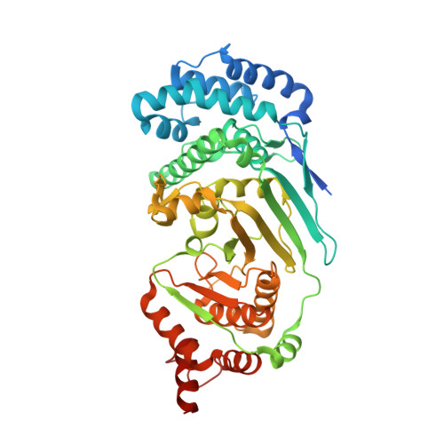
4EPP

Canonical poly(ADP-ribose) glycohydrolase from Tetrahymena thermophila.


Experimental Data Snapshot

  • Method: X-RAY DIFFRACTION
  • Resolution: 1.95 Å
  • R-Value Free: 0.207 
  • R-Value Work: 0.167 
  • R-Value Observed: 0.169 

wwPDB Validation   3D Report Full Report


Ligand Structure Quality Assessment 


This is version 1.1 of the entry. See complete history


Literature

Structure and mechanism of a canonical poly(ADP-ribose) glycohydrolase.

Dunstan, M.S.Barkauskaite, E.Lafite, P.Knezevic, C.E.Brassington, A.Ahel, M.Hergenrother, P.J.Leys, D.Ahel, I.

(2012) Nat Commun 3: 878-878

  • DOI: https://doi.org/10.1038/ncomms1889
  • Primary Citation of Related Structures:  
    4EPP, 4EPQ

  • PubMed Abstract: 

    Poly(ADP-ribosyl)ation is a reversible post-translational protein modification involved in the regulation of a number of cellular processes including DNA repair, chromatin structure, mitosis, transcription, checkpoint activation, apoptosis and asexual development. The reversion of poly(ADP-ribosyl)ation is catalysed by poly(ADP-ribose) (PAR) glycohydrolase (PARG), which specifically targets the unique PAR (1''-2') ribose-ribose bonds. Here we report the structure and mechanism of the first canonical PARG from the protozoan Tetrahymena thermophila. In addition, we reveal the structure of T. thermophila PARG in a complex with a novel rhodanine-containing mammalian PARG inhibitor RBPI-3. Our data demonstrate that the protozoan PARG represents a good model for human PARG and is therefore likely to prove useful in guiding structure-based discovery of new classes of PARG inhibitors.


  • Organizational Affiliation

    Manchester Interdisciplinary Biocentre, Princess Street 131, M1 7DN, Manchester, UK.


Macromolecules
Find similar proteins by:  (by identity cutoff)  |  3D Structure
Entity ID: 1
MoleculeChains Sequence LengthOrganismDetailsImage
Poly(ADP-ribose) glycohydrolase
A, B
477Tetrahymena thermophilaMutation(s): 0 
EC: 3.2.1.143
UniProt
Find proteins for I6L8L8 (Tetrahymena thermophila)
Explore I6L8L8 
Go to UniProtKB:  I6L8L8
Entity Groups  
Sequence Clusters30% Identity50% Identity70% Identity90% Identity95% Identity100% Identity
UniProt GroupI6L8L8
Sequence Annotations
Expand
  • Reference Sequence
Small Molecules
Ligands 1 Unique
IDChains Name / Formula / InChI Key2D Diagram3D Interactions
APR
Query on APR

Download Ideal Coordinates CCD File 
C [auth A],
D [auth B]
ADENOSINE-5-DIPHOSPHORIBOSE
C15 H23 N5 O14 P2
SRNWOUGRCWSEMX-KEOHHSTQSA-N
Experimental Data & Validation

Experimental Data

  • Method: X-RAY DIFFRACTION
  • Resolution: 1.95 Å
  • R-Value Free: 0.207 
  • R-Value Work: 0.167 
  • R-Value Observed: 0.169 
  • Space Group: P 41
Unit Cell:
Length ( Å )Angle ( ˚ )
a = 112.7α = 90
b = 112.7β = 90
c = 88.6γ = 90
Software Package:
Software NamePurpose
SOLVEphasing
PDB_EXTRACTdata extraction
ADSCdata collection
XDSdata reduction
XDSdata scaling
PHENIXrefinement

Structure Validation

View Full Validation Report


Ligand Structure Quality Assessment 


Entry History 

Deposition Data

Revision History  (Full details and data files)

  • Version 1.0: 2012-06-20
    Type: Initial release
  • Version 1.1: 2024-02-28
    Changes: Data collection, Database references, Derived calculations