4FIL

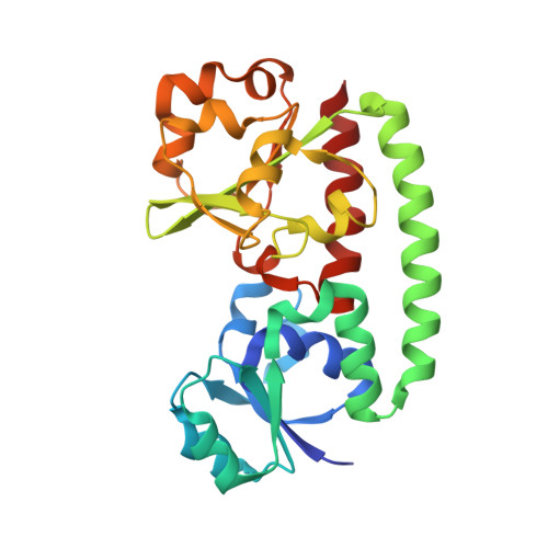
Structure of FhuD2 from Staphylococcus Aureus with Bound Ferrioxamine B


Experimental Data Snapshot

  • Method: X-RAY DIFFRACTION
  • Resolution: 2.40 Å
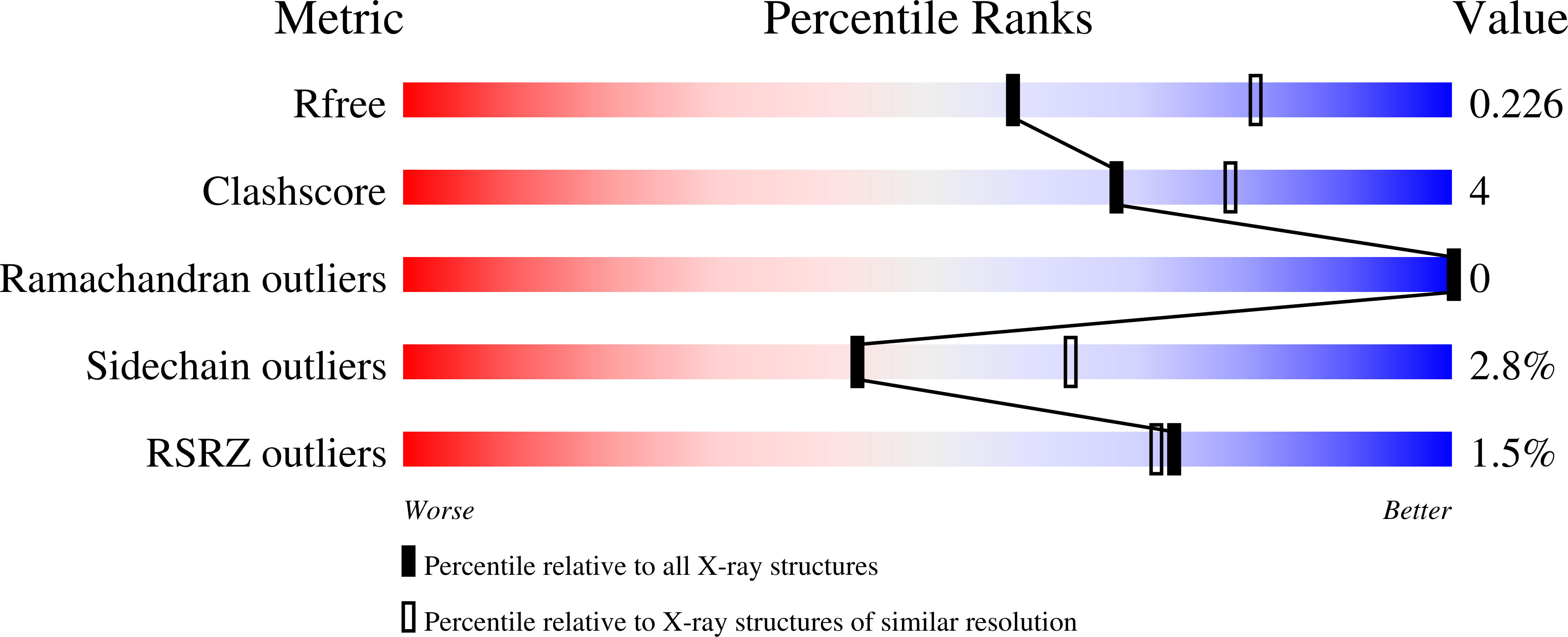
  • R-Value Free: 0.229 
  • R-Value Work: 0.198 
  • R-Value Observed: 0.201 

Starting Model: experimental
View more details

wwPDB Validation   3D Report Full Report


Ligand Structure Quality Assessment 


This is version 1.2 of the entry. See complete history


Literature

Crystal and solution structure analysis of FhuD2 from Staphylococcus aureus in multiple unliganded conformations and bound to ferrioxamine-B.

Podkowa, K.J.Briere, L.A.Heinrichs, D.E.Shilton, B.H.

(2014) Biochemistry 53: 2017-2031

  • DOI: https://doi.org/10.1021/bi401349d
  • Primary Citation of Related Structures:  
    4FIL, 4FKM, 4FNA

  • PubMed Abstract: 

    Iron acquisition is a central process for virtually all organisms. In Staphylococcus aureus, FhuD2 is a lipoprotein that is a high-affinity receptor for iron-bound hydroxamate siderophores. In this study, FhuD2 was crystallized bound to ferrioxamine-B (FXB), and also in its ligand-free state; the latter structures are the first for hydroxamate-binding receptors within this protein family. The structure of the FhuD2-FXB conformation shows that residues W197 and R199 from the C-terminal domain donate hydrogen bonds to the hydroxamate oxygens, and a ring of aromatic residues cradles the aliphatic arms connecting the hydroxamate moieties of the siderophore. The available ligand-bound structures of FhuD from Escherichia coli and YfiY from Bacillus cereus show that, despite a high degree of structural conservation, three protein families have evolved with critical siderophore binding residues on either the C-terminal domain (S. aureus), the N-terminal domain (E. coli), or both (B. cereus). Unliganded FhuD2 was crystallized in five conformations related by rigid body movements of the N- and C-terminal domains. Small-angle X-ray scattering (SAXS) indicates that the solution conformation of unliganded FhuD2 is more compact than the conformations observed in crystals. The ligand-induced conformational changes for FhuD2 in solution are relatively modest and depend on the identity of the siderophore. The crystallographic and SAXS results are used to discuss roles for the liganded and unliganded forms of FhuD2 in the siderophore transport mechanism.


  • Organizational Affiliation

    Department of Biochemistry and ‡Department of Microbiology and Immunology, The University of Western Ontario , London, Ontario, Canada N6A 5C1.


Macromolecules
Find similar proteins by:  (by identity cutoff)  |  3D Structure
Entity ID: 1
MoleculeChains Sequence LengthOrganismDetailsImage
Ferric hydroxamate receptor 2
A, B, C, D
261Staphylococcus aureusMutation(s): 3 
Gene Names: fhud2SAV2284
UniProt
Find proteins for A0A0H3JWU6 (Staphylococcus aureus (strain Mu50 / ATCC 700699))
Explore A0A0H3JWU6 
Go to UniProtKB:  A0A0H3JWU6
Entity Groups  
Sequence Clusters30% Identity50% Identity70% Identity90% Identity95% Identity100% Identity
UniProt GroupA0A0H3JWU6
Sequence Annotations
Expand
  • Reference Sequence
Small Molecules
Ligands 3 Unique
IDChains Name / Formula / InChI Key2D Diagram3D Interactions
0UE
Query on 0UE

Download Ideal Coordinates CCD File 
E [auth A],
FB [auth D],
SA [auth C],
Y [auth B]
Ferrioxamine B
C25 H45 Fe N6 O8
SRMBQCVUAVULDJ-UHFFFAOYSA-N
ZN
Query on ZN

Download Ideal Coordinates CCD File 
AA [auth B]
AB [auth C]
BA [auth B]
BB [auth C]
CA [auth B]
AA [auth B],
AB [auth C],
BA [auth B],
BB [auth C],
CA [auth B],
CB [auth C],
DA [auth B],
DB [auth C],
EA [auth B],
EB [auth C],
F [auth A],
FA [auth B],
G [auth A],
GA [auth B],
GB [auth D],
H [auth A],
HA [auth B],
HB [auth D],
I [auth A],
IA [auth B],
IB [auth D],
J [auth A],
JA [auth B],
JB [auth D],
K [auth A],
KA [auth B],
KB [auth D],
L [auth A],
LA [auth B],
LB [auth D],
M [auth A],
MA [auth B],
MB [auth D],
N [auth A],
NA [auth B],
NB [auth D],
O [auth A],
OA [auth B],
OB [auth D],
P [auth A],
PB [auth D],
Q [auth A],
QB [auth D],
R [auth A],
RB [auth D],
S [auth A],
SB [auth D],
TA [auth C],
TB [auth D],
UA [auth C],
VA [auth C],
WA [auth C],
XA [auth C],
YA [auth C],
Z [auth B],
ZA [auth C]
ZINC ION
Zn
PTFCDOFLOPIGGS-UHFFFAOYSA-N
EDO
Query on EDO

Download Ideal Coordinates CCD File 
PA [auth B]
QA [auth B]
RA [auth B]
T [auth A]
U [auth A]
PA [auth B],
QA [auth B],
RA [auth B],
T [auth A],
U [auth A],
UB [auth D],
V [auth A],
VB [auth D],
W [auth A],
WB [auth D],
X [auth A],
XB [auth D]
1,2-ETHANEDIOL
C2 H6 O2
LYCAIKOWRPUZTN-UHFFFAOYSA-N
Experimental Data & Validation

Experimental Data

  • Method: X-RAY DIFFRACTION
  • Resolution: 2.40 Å
  • R-Value Free: 0.229 
  • R-Value Work: 0.198 
  • R-Value Observed: 0.201 
  • Space Group: P 1 21 1
Unit Cell:
Length ( Å )Angle ( ˚ )
a = 81.343α = 90
b = 78.996β = 109.87
c = 116.811γ = 90
Software Package:
Software NamePurpose
ADSCdata collection
PHASERphasing
PHENIXrefinement
MOSFLMdata reduction
SCALAdata scaling

Structure Validation

View Full Validation Report


Ligand Structure Quality Assessment 


Entry History 

Deposition Data

Revision History  (Full details and data files)

  • Version 1.0: 2013-06-12
    Type: Initial release
  • Version 1.1: 2014-06-04
    Changes: Database references
  • Version 1.2: 2023-09-13
    Changes: Data collection, Database references, Derived calculations, Refinement description