4H54

Crystal structure of the diguanylate cyclase DgcZ


Experimental Data Snapshot

  • Method: X-RAY DIFFRACTION
  • Resolution: 3.90 Å
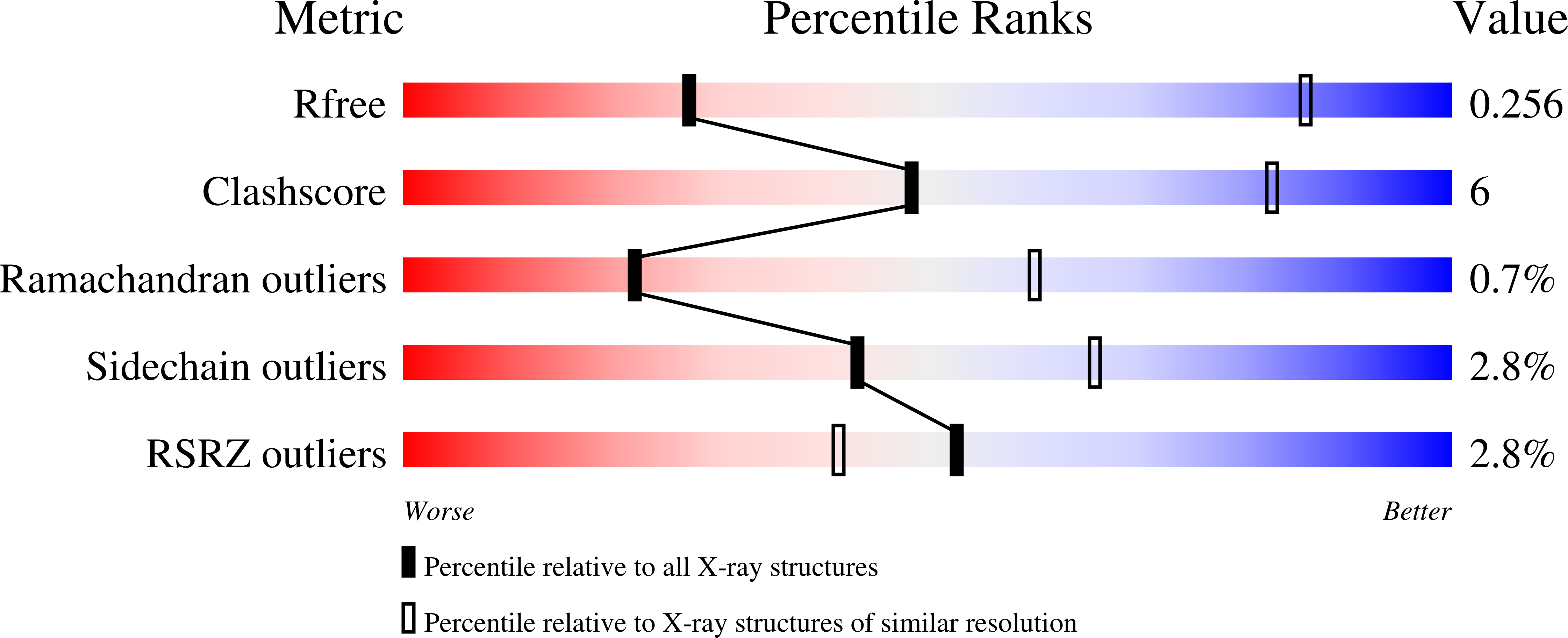
  • R-Value Free: 0.241 
  • R-Value Work: 0.242 
  • R-Value Observed: 0.242 

Starting Models: experimental
View more details

wwPDB Validation   3D Report Full Report


Ligand Structure Quality Assessment 


This is version 1.2 of the entry. See complete history


Literature

Structure and signaling mechanism of a zinc-sensory diguanylate cyclase.

Zahringer, F.Lacanna, E.Jenal, U.Schirmer, T.Boehm, A.

(2013) Structure 21: 1149-1157

  • DOI: https://doi.org/10.1016/j.str.2013.04.026
  • Primary Citation of Related Structures:  
    3T9O, 3TVK, 4H54

  • PubMed Abstract: 

    Diguanylate cyclases synthesize the second messenger c-di-GMP, which in turn governs a plethora of physiological processes in bacteria. Although most diguanylate cyclases harbor sensory domains, their input signals are largely unknown. Here, we demonstrate that diguanylate cyclase DgcZ (YdeH) from Escherichia coli is regulated allosterically by zinc. Crystal structures show that the zinc ion is bound to the 3His/1Cys motif of the regulatory chemoreceptor zinc-binding domain, which mediates subunit contact within the dimeric enzyme. In vitro, zinc reversibly inhibits DgcZ with a subfemtomolar Ki constant. In vivo, bacterial biofilm formation is modulated by externally applied zinc in a DgcZ- and c-di-GMP-dependent fashion. The study outlines the structural principles of this zinc sensor. Zinc binding seems to regulate the activity of the catalytic GGDEF domains by impeding their mobility and thus preventing productive encounter of the two GTP substrates.


  • Organizational Affiliation

    Focal Area of Structural Biology and Biophysics, Biozentrum, University of Basel, CH-4056 Basel, Switzerland.


Macromolecules
Find similar proteins by:  (by identity cutoff)  |  3D Structure
Entity ID: 1
MoleculeChains Sequence LengthOrganismDetailsImage
Diguanylate cyclase YdeH
A, B
304Escherichia coli K-12Mutation(s): 1 
Gene Names: b1535JW1528ydeGydeH
EC: 2.7.7.65
UniProt
Find proteins for P31129 (Escherichia coli (strain K12))
Explore P31129 
Go to UniProtKB:  P31129
Entity Groups  
Sequence Clusters30% Identity50% Identity70% Identity90% Identity95% Identity100% Identity
UniProt GroupP31129
Sequence Annotations
Expand
  • Reference Sequence
Small Molecules
Ligands 4 Unique
IDChains Name / Formula / InChI Key2D Diagram3D Interactions
C2E
Query on C2E

Download Ideal Coordinates CCD File 
G [auth A]9,9'-[(2R,3R,3aS,5S,7aR,9R,10R,10aS,12S,14aR)-3,5,10,12-tetrahydroxy-5,12-dioxidooctahydro-2H,7H-difuro[3,2-d:3',2'-j][1,3,7,9,2,8]tetraoxadiphosphacyclododecine-2,9-diyl]bis(2-amino-1,9-dihydro-6H-purin-6-one)
C20 H24 N10 O14 P2
PKFDLKSEZWEFGL-MHARETSRSA-N
GAV
Query on GAV

Download Ideal Coordinates CCD File 
D [auth A],
F [auth A],
I [auth B],
K [auth B]
GUANOSINE-5'-RP-ALPHA-THIO-TRIPHOSPHATE
C10 H16 N5 O13 P3 S
YRUWXBKVSXSSGS-KIOICMOXSA-N
ZN
Query on ZN

Download Ideal Coordinates CCD File 
C [auth A],
H [auth B]
ZINC ION
Zn
PTFCDOFLOPIGGS-UHFFFAOYSA-N
MG
Query on MG

Download Ideal Coordinates CCD File 
E [auth A],
J [auth B]
MAGNESIUM ION
Mg
JLVVSXFLKOJNIY-UHFFFAOYSA-N
Experimental Data & Validation

Experimental Data

  • Method: X-RAY DIFFRACTION
  • Resolution: 3.90 Å
  • R-Value Free: 0.241 
  • R-Value Work: 0.242 
  • R-Value Observed: 0.242 
  • Space Group: P 61
Unit Cell:
Length ( Å )Angle ( ˚ )
a = 102.593α = 90
b = 102.593β = 90
c = 129.156γ = 120
Software Package:
Software NamePurpose
REFMACrefinement
PDB_EXTRACTdata extraction
MARMOSAICdata collection
MOSFLMdata reduction
SCALAdata scaling
PHASERphasing
BUSTERrefinement

Structure Validation

View Full Validation Report


Ligand Structure Quality Assessment 


Entry History 

Deposition Data

Revision History  (Full details and data files)

  • Version 1.0: 2013-07-03
    Type: Initial release
  • Version 1.1: 2013-07-17
    Changes: Database references
  • Version 1.2: 2023-09-20
    Changes: Data collection, Database references, Derived calculations, Refinement description, Structure summary