4HB2

Crystal structure of CRM1-Ran-RanBP1


Experimental Data Snapshot

  • Method: X-RAY DIFFRACTION
  • Resolution: 1.80 Å
  • R-Value Free: 0.201 
  • R-Value Work: 0.155 
  • R-Value Observed: 0.157 

wwPDB Validation   3D Report Full Report


Ligand Structure Quality Assessment 


This is version 1.4 of the entry. See complete history


Literature

Nuclear export inhibition through covalent conjugation and hydrolysis of Leptomycin B by CRM1.

Sun, Q.Carrasco, Y.P.Hu, Y.Guo, X.Mirzaei, H.Macmillan, J.Chook, Y.M.

(2013) Proc Natl Acad Sci U S A 110: 1303-1308

  • DOI: https://doi.org/10.1073/pnas.1217203110
  • Primary Citation of Related Structures:  
    4HAT, 4HAU, 4HAV, 4HAW, 4HAX, 4HAY, 4HAZ, 4HB0, 4HB2, 4HB3, 4HB4

  • PubMed Abstract: 

    The polyketide natural product Leptomycin B inhibits nuclear export mediated by the karyopherin protein chromosomal region maintenance 1 (CRM1). Here, we present 1.8- to 2.0-Å-resolution crystal structures of CRM1 bound to Leptomycin B and related inhibitors Anguinomycin A and Ratjadone A. Structural and complementary chemical analyses reveal an unexpected mechanism of inhibition involving covalent conjugation and CRM1-mediated hydrolysis of the natural products' lactone rings. Furthermore, mutagenesis reveals the mechanism of hydrolysis by CRM1. The nuclear export signal (NES)-binding groove of CRM1 is able to drive a chemical reaction in addition to binding protein cargoes for transport through the nuclear pore complex.


  • Organizational Affiliation

    Department of Pharmacology, University of Texas Southwestern Medical Center, Dallas, TX 75390-9041, USA.


Macromolecules
Find similar proteins by:  (by identity cutoff)  |  3D Structure
Entity ID: 1
MoleculeChains Sequence LengthOrganismDetailsImage
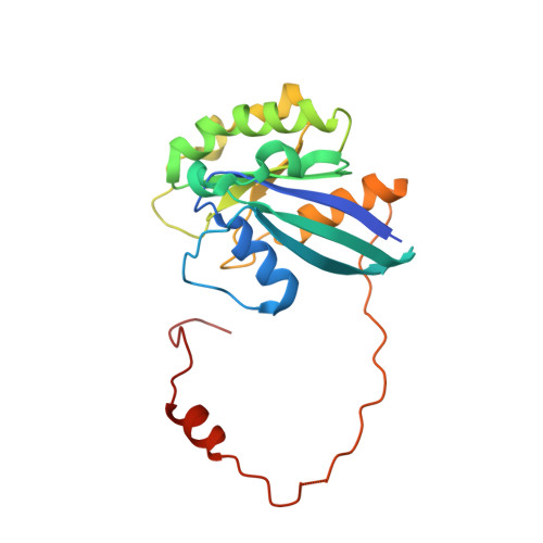
GTP-binding nuclear protein Ran216Homo sapiensMutation(s): 0 
Gene Names: RANARA24OK/SW-cl.81
EC: 3.6.5
UniProt & NIH Common Fund Data Resources
Find proteins for P62826 (Homo sapiens)
Explore P62826 
Go to UniProtKB:  P62826
PHAROS:  P62826
GTEx:  ENSG00000132341 
Entity Groups  
Sequence Clusters30% Identity50% Identity70% Identity90% Identity95% Identity100% Identity
UniProt GroupP62826
Sequence Annotations
Expand
  • Reference Sequence
Find similar proteins by:  (by identity cutoff)  |  3D Structure
Entity ID: 2
MoleculeChains Sequence LengthOrganismDetailsImage
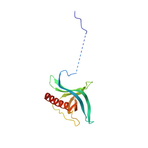
Ran-specific GTPase-activating protein 1140Saccharomyces cerevisiaeMutation(s): 0 
Gene Names: YRB1CST20HTN1SFO1YDR002WYD8119.08
UniProt
Find proteins for P41920 (Saccharomyces cerevisiae (strain ATCC 204508 / S288c))
Explore P41920 
Go to UniProtKB:  P41920
Entity Groups  
Sequence Clusters30% Identity50% Identity70% Identity90% Identity95% Identity100% Identity
UniProt GroupP41920
Sequence Annotations
Expand
  • Reference Sequence
Find similar proteins by:  (by identity cutoff)  |  3D Structure
Entity ID: 3
MoleculeChains Sequence LengthOrganismDetailsImage
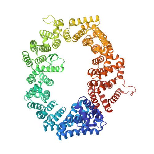
Exportin-11,023Saccharomyces cerevisiaeMutation(s): 2 
Gene Names: CRM1KAP124XPO1YGR218WG8514
UniProt
Find proteins for P30822 (Saccharomyces cerevisiae (strain ATCC 204508 / S288c))
Explore P30822 
Go to UniProtKB:  P30822
Entity Groups  
Sequence Clusters30% Identity50% Identity70% Identity90% Identity95% Identity100% Identity
UniProt GroupP30822
Sequence Annotations
Expand
  • Reference Sequence
Small Molecules
Ligands 5 Unique
IDChains Name / Formula / InChI Key2D Diagram3D Interactions
GNP
Query on GNP

Download Ideal Coordinates CCD File 
D [auth A]PHOSPHOAMINOPHOSPHONIC ACID-GUANYLATE ESTER
C10 H17 N6 O13 P3
UQABYHGXWYXDTK-UUOKFMHZSA-N
GOL
Query on GOL

Download Ideal Coordinates CCD File 
F [auth A],
I [auth C],
J [auth C],
O [auth C]
GLYCEROL
C3 H8 O3
PEDCQBHIVMGVHV-UHFFFAOYSA-N
EDO
Query on EDO

Download Ideal Coordinates CCD File 
G [auth A],
K [auth C]
1,2-ETHANEDIOL
C2 H6 O2
LYCAIKOWRPUZTN-UHFFFAOYSA-N
CL
Query on CL

Download Ideal Coordinates CCD File 
H [auth A],
L [auth C],
M [auth C],
N [auth C]
CHLORIDE ION
Cl
VEXZGXHMUGYJMC-UHFFFAOYSA-M
MG
Query on MG

Download Ideal Coordinates CCD File 
E [auth A]MAGNESIUM ION
Mg
JLVVSXFLKOJNIY-UHFFFAOYSA-N
Experimental Data & Validation

Experimental Data

  • Method: X-RAY DIFFRACTION
  • Resolution: 1.80 Å
  • R-Value Free: 0.201 
  • R-Value Work: 0.155 
  • R-Value Observed: 0.157 
  • Space Group: P 43 21 2
Unit Cell:
Length ( Å )Angle ( ˚ )
a = 106.328α = 90
b = 106.328β = 90
c = 306.69γ = 90
Software Package:
Software NamePurpose
REFMACrefinement

Structure Validation

View Full Validation Report


Ligand Structure Quality Assessment 


Entry History 

Deposition Data

Revision History  (Full details and data files)

  • Version 1.0: 2013-01-09
    Type: Initial release
  • Version 1.1: 2013-01-23
    Changes: Database references
  • Version 1.2: 2013-02-06
    Changes: Database references
  • Version 1.3: 2017-07-26
    Changes: Source and taxonomy
  • Version 1.4: 2024-02-28
    Changes: Data collection, Database references, Derived calculations