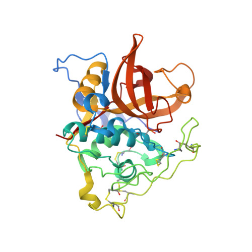
4I05

Structure of intermediate processing form of cathepsin B1 from Schistosoma mansoni


Experimental Data Snapshot

  • Method: X-RAY DIFFRACTION
  • Resolution: 1.90 Å
  • R-Value Free: 0.216 
  • R-Value Work: 0.163 
  • R-Value Observed: 0.166 

Starting Model: experimental
View more details

wwPDB Validation   3D Report Full Report


This is version 1.4 of the entry. See complete history


Literature

Activation route of the Schistosoma mansoni cathepsin B1 drug target: structural map with a glycosaminoglycan switch

Jilkova, A.Horn, M.Rezacova, P.Maresova, L.Fajtova, P.Brynda, J.Vondrasek, J.McKerrow, J.H.Caffrey, C.R.Mares, M.

(2014) Structure 22: 1786-1798

  • DOI: https://doi.org/10.1016/j.str.2014.09.015
  • Primary Citation of Related Structures:  
    4I04, 4I05, 4I07

  • PubMed Abstract: 

    Cathepsin B1 (SmCB1) is a digestive protease of the parasitic blood fluke Schistosoma mansoni and a drug target for the treatment of schistosomiasis, a disease that afflicts over 200 million people. SmCB1 is synthesized as an inactive zymogen in which the N-terminal propeptide blocks the active site. We investigated the activation of the zymogen by which the propeptide is proteolytically removed and its regulation by sulfated polysaccharides (SPs). We determined crystal structures of three molecular forms of SmCB1 along the activation pathway: the zymogen, an activation intermediate with a partially cleaved propeptide, and the mature enzyme. We demonstrate that SPs are essential for the autocatalytic activation of SmCB1, as they interact with a specific heparin-binding domain in the propeptide. An alternative activation route is mediated by an S. mansoni asparaginyl endopeptidase (legumain) which is downregulated by SPs, indicating that SPs act as a molecular switch between both activation mechanisms.


  • Organizational Affiliation

    Institute of Organic Chemistry and Biochemistry, Academy of Sciences of the Czech Republic, 16610 Prague, Czech Republic; Department of Biochemistry, Faculty of Science, Charles University in Prague, 12843 Prague, Czech Republic.


Macromolecules
Find similar proteins by:  (by identity cutoff)  |  3D Structure
Entity ID: 1
MoleculeChains Sequence LengthOrganismDetailsImage
Cathepsin B-like peptidase (C01 family)285Schistosoma mansoniMutation(s): 2 
Gene Names: cb1.1Smp_103610
EC: 3.4.22.1
UniProt
Find proteins for Q8MNY2 (Schistosoma mansoni)
Explore Q8MNY2 
Go to UniProtKB:  Q8MNY2
Entity Groups  
Sequence Clusters30% Identity50% Identity70% Identity90% Identity95% Identity100% Identity
UniProt GroupQ8MNY2
Sequence Annotations
Expand
  • Reference Sequence
Small Molecules
Ligands 1 Unique
IDChains Name / Formula / InChI Key2D Diagram3D Interactions
NA
Query on NA

Download Ideal Coordinates CCD File 
B [auth A]SODIUM ION
Na
FKNQFGJONOIPTF-UHFFFAOYSA-N
Modified Residues  1 Unique
IDChains TypeFormula2D DiagramParent
CSO
Query on CSO
A
L-PEPTIDE LINKINGC3 H7 N O3 SCYS
Experimental Data & Validation

Experimental Data

  • Method: X-RAY DIFFRACTION
  • Resolution: 1.90 Å
  • R-Value Free: 0.216 
  • R-Value Work: 0.163 
  • R-Value Observed: 0.166 
  • Space Group: C 1 2 1
Unit Cell:
Length ( Å )Angle ( ˚ )
a = 93.859α = 90
b = 50.988β = 91.31
c = 62.546γ = 90
Software Package:
Software NamePurpose
HKL-3000data collection
MOLREPphasing
REFMACrefinement
HKL-3000data reduction
HKL-3000data scaling

Structure Validation

View Full Validation Report


Entry History 

Deposition Data

Revision History  (Full details and data files)

  • Version 1.0: 2014-02-05
    Type: Initial release
  • Version 1.1: 2016-05-25
    Changes: Database references
  • Version 1.2: 2023-11-08
    Changes: Data collection, Database references, Derived calculations, Refinement description
  • Version 1.3: 2023-12-06
    Changes: Data collection
  • Version 1.4: 2024-10-16
    Changes: Structure summary