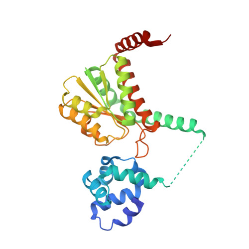
Structural insights into the regulation of sialic acid catabolism by the Vibrio vulnificus transcriptional repressor NanR
Hwang, J., Kim, B.S., Jang, S.Y., Lim, J.G., You, D.J., Jung, H.S., Oh, T.K., Lee, J.O., Choi, S.H., Kim, M.H.(2013) Proc Natl Acad Sci U S A 110: E2829-E2837
- PubMed: 23832782
- DOI: https://doi.org/10.1073/pnas.1302859110
- Primary Citation of Related Structures:
4IVN - PubMed Abstract:
Pathogenic and commensal bacteria that experience limited nutrient availability in their host have evolved sophisticated systems to catabolize the mucin sugar N-acetylneuraminic acid, thereby facilitating their survival and colonization. The correct function of the associated catabolic machinery is particularly crucial for the pathogenesis of enteropathogenic bacteria during infection, although the molecular mechanisms involved with the regulation of the catabolic machinery are unknown. This study reports the complex structure of NanR, a repressor of the N-acetylneuraminate (nan) genes responsible for N-acetylneuraminic acid catabolism, and its regulatory ligand, N-acetylmannosamine 6-phosphate (ManNAc-6P), in the human pathogenic bacterium Vibrio vulnificus. Structural studies combined with electron microscopic, biochemical, and in vivo analysis demonstrated that NanR forms a dimer in which the two monomers create an arched tunnel-like DNA-binding space, which contains positively charged residues that interact with the nan promoter. The interaction between the NanR dimer and DNA is alleviated by the ManNAc-6P-mediated relocation of residues in the ligand-binding domain of NanR, which subsequently relieves the repressive effect of NanR and induces the transcription of the nan genes. Survival studies in which mice were challenged with a ManNAc-6P-binding-defective mutant strain of V. vulnificus demonstrated that this relocation of NanR residues is critical for V. vulnificus pathogenesis. In summary, this study presents a model of the mechanism that regulates sialic acid catabolism via NanR in V. vulnificus.
- Infection and Immunity Research Center, Korea Research Institute of Bioscience and Biotechnology, Daejeon 305-806, Korea.
Organizational Affiliation:


