4K43

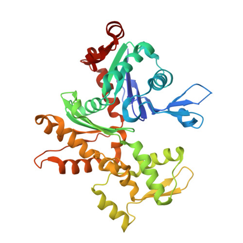
Crystal structure of actin in complex with synthetic AplC tail analogue GC04 [N-{(1E,4R,5R,7E,9S,10S,11S)-4,10-dimethoxy-11-[(2S,4S,5S)-2-(4-methoxyphenyl)-5-methyl-1,3-dioxan-4-yl]-5,9-dimethyl-6-oxododeca-1,7-dien-1-yl}-N-methylformamide]


Experimental Data Snapshot

  • Method: X-RAY DIFFRACTION
  • Resolution: 2.90 Å
  • R-Value Free: 0.277 
  • R-Value Work: 0.222 
  • R-Value Observed: 0.225 

wwPDB Validation   3D Report Full Report


Ligand Structure Quality Assessment 


This is version 1.2 of the entry. See complete history


Literature

Structural and biochemical studies of actin in complex with synthetic macrolide tail analogues.

Pereira, J.H.Petchprayoon, C.Hoepker, A.C.Moriarty, N.W.Fink, S.J.Cecere, G.Paterson, I.Adams, P.D.Marriott, G.

(2014) ChemMedChem 9: 2286-2293

  • DOI: https://doi.org/10.1002/cmdc.201402150
  • Primary Citation of Related Structures:  
    4K41, 4K42, 4K43

  • PubMed Abstract: 

    The actin filament-binding and filament-severing activities of the aplyronine, kabiramide, and reidispongiolide families of marine macrolides are located within the hydrophobic tail region of the molecule. Two synthetic tail analogues of aplyronine C (SF-01 and GC-04) are shown to bind to G-actin with dissociation constants of (285±33) and (132±13) nM, respectively. The crystal structures of actin complexes with GC-04, SF-01, and kabiramide C reveal a conserved mode of tail binding within the cleft that forms between subdomains (SD) 1 and 3. Our studies support the view that filament severing is brought about by specific binding of the tail region to the SD1/SD3 cleft on the upper protomer, which displaces loop-D from the lower protomer on the same half-filament. With previous studies showing that the GC-04 analogue can sever actin filaments, it is argued that the shorter complex lifetime of tail analogues with F-actin would make them more effective at severing filaments compared with plasma gelsolin. Structure-based analyses are used to suggest more reactive or targetable forms of GC-04 and SF-01, which may serve to boost the capacity of the serum actin scavenging system, to generate antibody conjugates against tumor cell antigens, and to decrease sputum viscosity in children with cystic fibrosis.


  • Organizational Affiliation

    Physical Biosciences Division, Lawrence Berkeley National Laboratory, Berkeley, CA 94720 (USA).


Macromolecules
Find similar proteins by:  (by identity cutoff)  |  3D Structure
Entity ID: 1
MoleculeChains Sequence LengthOrganismDetailsImage
Actin, alpha skeletal muscle
A, B
375Oryctolagus cuniculusMutation(s): 0 
EC: 3.6.4
UniProt
Find proteins for P68135 (Oryctolagus cuniculus)
Explore P68135 
Go to UniProtKB:  P68135
Entity Groups  
Sequence Clusters30% Identity50% Identity70% Identity90% Identity95% Identity100% Identity
UniProt GroupP68135
Sequence Annotations
Expand
  • Reference Sequence
Small Molecules
Ligands 3 Unique
IDChains Name / Formula / InChI Key2D Diagram3D Interactions
1PO
Query on 1PO

Download Ideal Coordinates CCD File 
E [auth A],
H [auth B]
N-{(1E,4R,5R,7E,9S,10S,11S)-4,10-dimethoxy-11-[(2S,4S,5S)-2-(4-methoxyphenyl)-5-methyl-1,3-dioxan-4-yl]-5,9-dimethyl-6-oxododeca-1,7-dien-1-yl}-N-methylformamide
C30 H45 N O7
NMVATSAEADBXDC-AUYAIVLXSA-N
ADP
Query on ADP

Download Ideal Coordinates CCD File 
D [auth A],
G [auth B]
ADENOSINE-5'-DIPHOSPHATE
C10 H15 N5 O10 P2
XTWYTFMLZFPYCI-KQYNXXCUSA-N
CA
Query on CA

Download Ideal Coordinates CCD File 
C [auth A],
F [auth B]
CALCIUM ION
Ca
BHPQYMZQTOCNFJ-UHFFFAOYSA-N
Experimental Data & Validation

Experimental Data

  • Method: X-RAY DIFFRACTION
  • Resolution: 2.90 Å
  • R-Value Free: 0.277 
  • R-Value Work: 0.222 
  • R-Value Observed: 0.225 
  • Space Group: P 1 21 1
Unit Cell:
Length ( Å )Angle ( ˚ )
a = 54.578α = 90
b = 70.349β = 104.7
c = 107.673γ = 90
Software Package:
Software NamePurpose
HKL-2000data collection
PHASERphasing
PHENIXrefinement
HKL-2000data reduction
HKL-2000data scaling

Structure Validation

View Full Validation Report


Ligand Structure Quality Assessment 


Entry History 

Deposition Data

Revision History  (Full details and data files)

  • Version 1.0: 2014-10-01
    Type: Initial release
  • Version 1.1: 2014-10-08
    Changes: Database references
  • Version 1.2: 2024-02-28
    Changes: Data collection, Database references, Derived calculations