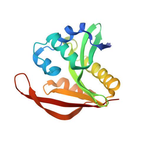
4KLV

Crystal structure of a GNAT superfamily acetyltransferase PA4794 in complex with 4-methylumbelliferyl phosphate


Experimental Data Snapshot

  • Method: X-RAY DIFFRACTION
  • Resolution: 1.30 Å
  • R-Value Free: 0.152 
  • R-Value Work: 0.122 
  • R-Value Observed: 0.124 

Starting Model: experimental
View more details

wwPDB Validation   3D Report Full Report


Ligand Structure Quality Assessment 


This is version 1.4 of the entry. See complete history


Literature

Structural, Functional, and Inhibition Studies of a Gcn5-related N-Acetyltransferase (GNAT) Superfamily Protein PA4794: A NEW C-TERMINAL LYSINE PROTEIN ACETYLTRANSFERASE FROM PSEUDOMONAS AERUGINOSA.

Majorek, K.A.Kuhn, M.L.Chruszcz, M.Anderson, W.F.Minor, W.

(2013) J Biol Chem 288: 30223-30235

  • DOI: https://doi.org/10.1074/jbc.M113.501353
  • Primary Citation of Related Structures:  
    3PGP, 4KLV, 4KLW, 4KOR, 4KOS, 4KOT, 4KOU, 4KOV, 4KOW, 4KOX, 4KOY, 4KUA, 4KUB, 4L89, 4L8A

  • PubMed Abstract: 

    The Gcn5-related N-acetyltransferase (GNAT) superfamily is a large group of evolutionarily related acetyltransferases, with multiple paralogs in organisms from all kingdoms of life. The functionally characterized GNATs have been shown to catalyze the transfer of an acetyl group from acetyl-coenzyme A (Ac-CoA) to the amine of a wide range of substrates, including small molecules and proteins. GNATs are prevalent and implicated in a myriad of aspects of eukaryotic and prokaryotic physiology, but functions of many GNATs remain unknown. In this work, we used a multi-pronged approach of x-ray crystallography and biochemical characterization to elucidate the sequence-structure-function relationship of the GNAT superfamily member PA4794 from Pseudomonas aeruginosa. We determined that PA4794 acetylates the Nε amine of a C-terminal lysine residue of a peptide, suggesting it is a protein acetyltransferase specific for a C-terminal lysine of a substrate protein or proteins. Furthermore, we identified a number of molecules, including cephalosporin antibiotics, which are inhibitors of PA4794 and bind in its substrate-binding site. Often, these molecules mimic the conformation of the acetylated peptide product. We have determined structures of PA4794 in the apo-form, in complexes with Ac-CoA, CoA, several antibiotics and other small molecules, and a ternary complex with the products of the reaction: CoA and acetylated peptide. Also, we analyzed PA4794 mutants to identify residues important for substrate binding and catalysis.


  • Organizational Affiliation

    From the Department of Molecular Physiology and Biological Physics, University of Virginia, Charlottesville, Virginia 22908,; the Bioinformatics Laboratory, Institute of Molecular Biology and Biotechnology, Faculty of Biology, Adam Mickiewicz University, 61-614 Poznan, Poland,; the Midwest Center for Structural Genomics, Argonne National Laboratory, Argonne, Illinois 60439, and; the Center for Structural Genomics of Infectious Diseases (CSGID).


Macromolecules
Find similar proteins by:  (by identity cutoff)  |  3D Structure
Entity ID: 1
MoleculeChains Sequence LengthOrganismDetailsImage
Uncharacterized protein162Pseudomonas aeruginosa PAO1Mutation(s): 0 
Gene Names: PA4794
UniProt
Find proteins for Q9HV14 (Pseudomonas aeruginosa (strain ATCC 15692 / DSM 22644 / CIP 104116 / JCM 14847 / LMG 12228 / 1C / PRS 101 / PAO1))
Explore Q9HV14 
Go to UniProtKB:  Q9HV14
Entity Groups  
Sequence Clusters30% Identity50% Identity70% Identity90% Identity95% Identity100% Identity
UniProt GroupQ9HV14
Sequence Annotations
Expand
  • Reference Sequence
Small Molecules
Ligands 3 Unique
IDChains Name / Formula / InChI Key2D Diagram3D Interactions
KLV
Query on KLV

Download Ideal Coordinates CCD File 
Q [auth A]4-methylumbelliferyl phosphate
C10 H9 O6 P
BCHIXGBGRHLSBE-UHFFFAOYSA-N
SO4
Query on SO4

Download Ideal Coordinates CCD File 
B [auth A]
C [auth A]
D [auth A]
E [auth A]
F [auth A]
B [auth A],
C [auth A],
D [auth A],
E [auth A],
F [auth A],
L [auth A],
M [auth A]
SULFATE ION
O4 S
QAOWNCQODCNURD-UHFFFAOYSA-L
EDO
Query on EDO

Download Ideal Coordinates CCD File 
G [auth A]
H [auth A]
I [auth A]
J [auth A]
K [auth A]
G [auth A],
H [auth A],
I [auth A],
J [auth A],
K [auth A],
N [auth A],
O [auth A],
P [auth A],
R [auth A],
S [auth A],
T [auth A]
1,2-ETHANEDIOL
C2 H6 O2
LYCAIKOWRPUZTN-UHFFFAOYSA-N
Binding Affinity Annotations 
IDSourceBinding Affinity
KLV PDBBind:  4KLV Kd: 1.65e+5 (nM) from 1 assay(s)
Experimental Data & Validation

Experimental Data

  • Method: X-RAY DIFFRACTION
  • Resolution: 1.30 Å
  • R-Value Free: 0.152 
  • R-Value Work: 0.122 
  • R-Value Observed: 0.124 
  • Space Group: P 21 21 2
Unit Cell:
Length ( Å )Angle ( ˚ )
a = 57.34α = 90
b = 76.486β = 90
c = 39.336γ = 90
Software Package:
Software NamePurpose
HKL-3000data collection
HKL-3000phasing
REFMACrefinement
Cootmodel building
HKL-3000data reduction
HKL-3000data scaling

Structure Validation

View Full Validation Report


Ligand Structure Quality Assessment 


Entry History 

Deposition Data

Revision History  (Full details and data files)

  • Version 1.0: 2013-05-22
    Type: Initial release
  • Version 1.1: 2013-09-18
    Changes: Database references
  • Version 1.2: 2013-11-13
    Changes: Database references
  • Version 1.3: 2022-04-13
    Changes: Database references, Derived calculations, Structure summary
  • Version 1.4: 2023-09-20
    Changes: Data collection, Refinement description