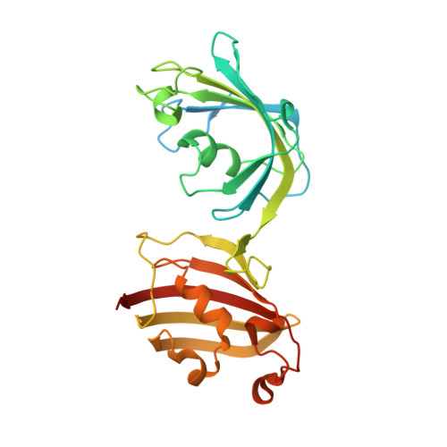
4LAY

Crystal Structure Analysis of FKBP52, Complex with I63


Experimental Data Snapshot

  • Method: X-RAY DIFFRACTION
  • Resolution: 1.70 Å
  • R-Value Free: 0.205 
  • R-Value Work: 0.168 
  • R-Value Observed: 0.170 

Starting Model: experimental
View more details

wwPDB Validation   3D Report Full Report


Ligand Structure Quality Assessment 


This is version 1.3 of the entry. See complete history


Literature

Crystal Structures of the Free and Ligand-Bound FK1-FK2 Domain Segment of FKBP52 Reveal a Flexible Inter-Domain Hinge.

Bracher, A.Kozany, C.Hahle, A.Wild, P.Zacharias, M.Hausch, F.

(2013) J Mol Biol 425: 4134-4144

  • DOI: https://doi.org/10.1016/j.jmb.2013.07.041
  • Primary Citation of Related Structures:  
    4LAV, 4LAW, 4LAX, 4LAY

  • PubMed Abstract: 

    The human Hsp90 co-chaperone FKBP52 belongs to the family of FK506-binding proteins, which act as peptidyl-prolyl isomerases. FKBP52 specifically enhances the signaling of steroid hormone receptors, modulates ion channels and regulates neuronal outgrowth dynamics. In turn, small-molecule ligands of FKBP52 have been suggested as potential neurotrophic or anti-prostate cancer agents. The usefulness of available ligands is however limited by a lack of selectivity. The immunophilin FKBP52 is composed of three domains, an FK506-binding domain with peptidyl-prolyl isomerase activity, an FKBP-like domain of unknown function and a TPR-clamp domain, which recognizes the C-terminal peptide of Hsp90 with high affinity. The herein reported crystal structures of FKBP52 reveal that the short linker connecting the FK506-binding domain and the FKBP-like domain acts as a flexible hinge. This enhanced flexibility and its modulation by phosphorylation might explain some of the functional antagonism between the closely related homologs FKBP51 and FKBP52. We further present two co-crystal structures of FKBP52 in complex with the prototypic ligand FK506 and a synthetic analog thereof. These structures revealed the molecular interactions in great detail, which enabled in-depth comparison with the corresponding complexes of the other cytosolic FKBPs, FKBP51 and FKBP12. The observed subtle differences provide crucial insights for the rational design of ligands with improved selectivity for FKBP52.


  • Organizational Affiliation

    Department of Cellular Biochemistry, Max Planck Institute of Biochemistry, Am Klopferspitz 18, 82152 Martinsried, Germany. Electronic address: bracher@biochem.mpg.de.


Macromolecules
Find similar proteins by:  (by identity cutoff)  |  3D Structure
Entity ID: 1
MoleculeChains Sequence LengthOrganismDetailsImage
Peptidyl-prolyl cis-trans isomerase FKBP4280Homo sapiensMutation(s): 0 
Gene Names: FKBP4FKBP52
EC: 5.2.1.8
UniProt & NIH Common Fund Data Resources
Find proteins for Q02790 (Homo sapiens)
Explore Q02790 
Go to UniProtKB:  Q02790
PHAROS:  Q02790
GTEx:  ENSG00000004478 
Entity Groups  
Sequence Clusters30% Identity50% Identity70% Identity90% Identity95% Identity100% Identity
UniProt GroupQ02790
Sequence Annotations
Expand
  • Reference Sequence
Small Molecules
Ligands 1 Unique
IDChains Name / Formula / InChI Key2D Diagram3D Interactions
I63
Query on I63

Download Ideal Coordinates CCD File 
B [auth A]{3-[(1R)-3-(3,4-dimethoxyphenyl)-1-({[(2S)-1-(3,3-dimethyl-2-oxopentanoyl)piperidin-2-yl]carbonyl}oxy)propyl]phenoxy}acetic acid
C32 H41 N O9
CKUAMXWZIHXZJC-LOSJGSFVSA-N
Binding Affinity Annotations 
IDSourceBinding Affinity
I63 BindingDB:  4LAY Ki: 20 (nM) from 1 assay(s)
IC50: min: 9370, max: 1.05e+4 (nM) from 2 assay(s)
Experimental Data & Validation

Experimental Data

  • Method: X-RAY DIFFRACTION
  • Resolution: 1.70 Å
  • R-Value Free: 0.205 
  • R-Value Work: 0.168 
  • R-Value Observed: 0.170 
  • Space Group: P 1 21 1
Unit Cell:
Length ( Å )Angle ( ˚ )
a = 47.592α = 90
b = 42.351β = 105.75
c = 76.811γ = 90
Software Package:
Software NamePurpose
MOSFLMdata reduction
SCALAdata scaling
MOLREPphasing
REFMACrefinement
PDB_EXTRACTdata extraction

Structure Validation

View Full Validation Report


Ligand Structure Quality Assessment 


Entry History 

Deposition Data

Revision History  (Full details and data files)

  • Version 1.0: 2013-08-21
    Type: Initial release
  • Version 1.1: 2013-08-28
    Changes: Database references
  • Version 1.2: 2013-11-13
    Changes: Database references
  • Version 1.3: 2023-09-20
    Changes: Data collection, Database references, Derived calculations, Refinement description