4LXR | pdb_00004lxr

Structure of the Toll - Spatzle complex, a molecular hub in Drosophila development and innate immunity


Experimental Data Snapshot

  • Method: X-RAY DIFFRACTION
  • Resolution: 2.20 Å
  • R-Value Free: 
    0.218 (Depositor), 0.218 (DCC) 
  • R-Value Work: 
    0.176 (Depositor), 0.178 (DCC) 
  • R-Value Observed: 
    0.178 (Depositor) 

wwPDB Validation   3D Report Full Report


Ligand Structure Quality Assessment 

Created with Raphaël 2.3.0Worse 01 BetterLigand structure goodness of fit to experimental dataBest fitted NAGClick on this verticalbar to view details

This is version 2.1 of the entry. See complete history


Literature

Structure of the Toll-Spatzle complex, a molecular hub in Drosophila development and innate immunity.

Parthier, C.Stelter, M.Ursel, C.Fandrich, U.Lilie, H.Breithaupt, C.Stubbs, M.T.

(2014) Proc Natl Acad Sci U S A 111: 6281-6286

  • DOI: https://doi.org/10.1073/pnas.1320678111
  • Primary Citation of Related Structures:  
    4LXR, 4LXS

  • PubMed Abstract: 

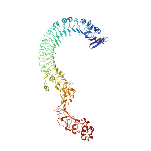
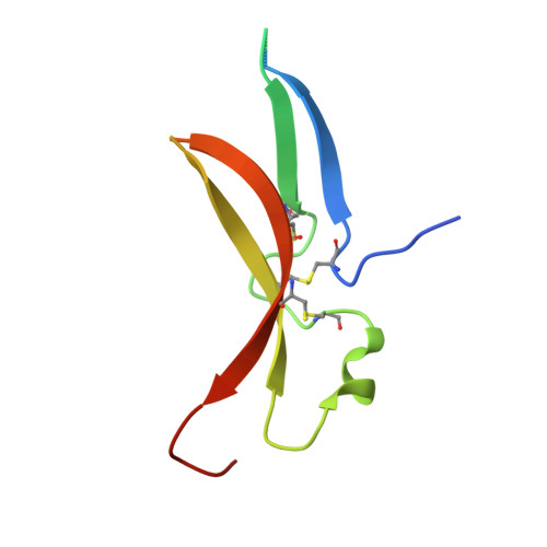
    Drosophila Toll receptors are involved in embryonic development and the immune response of adult flies. In both processes, the only known Toll receptor ligand is the human nerve growth factor-like cystine knot protein Spätzle. Here we present the crystal structure of a 1:1 (nonsignaling) complex of the full-length Toll receptor ectodomain (ECD) with the Spätzle cystine knot domain dimer. The ECD is divided into two leucine-rich repeat (LRR) domains, each of which is capped by cysteine-rich domains. Spätzle binds to the concave surface of the membrane-distal LRR domain, in contrast to the flanking ligand interactions observed for mammalian Toll-like receptors, with asymmetric contributions from each Spätzle protomer. The structure allows rationalization of existing genetic and biochemical data and provides a framework for targeting the immune systems of insects of economic importance, as well as a variety of invertebrate disease vectors.


  • Organizational Affiliation

    Institut für Biochemie und Biotechnologie and Mitteldeutsches Zentrum für Struktur und Dynamik von Proteinen, Martin-Luther-Universität Halle-Wittenberg, 06120 Halle (Saale), Germany.


Macromolecules
Find similar proteins by:  (by identity cutoff)  |  3D Structure
Entity ID: 1
MoleculeChains Sequence LengthOrganismDetailsImage
Protein toll783Drosophila melanogasterMutation(s): 0 
Gene Names: TlCG5490
UniProt
Find proteins for P08953 (Drosophila melanogaster)
Explore P08953 
Go to UniProtKB:  P08953
Entity Groups  
Sequence Clusters30% Identity50% Identity70% Identity90% Identity95% Identity100% Identity
UniProt GroupP08953
Glycosylation
Glycosylation Sites: 10Go to GlyGen: P08953-1
Sequence Annotations
Expand
  • Reference Sequence
Find similar proteins by:  (by identity cutoff)  |  3D Structure
Entity ID: 2
MoleculeChains Sequence LengthOrganismDetailsImage
Protein spaetzle C-106B [auth J],
C [auth K]
114Drosophila melanogasterMutation(s): 0 
Gene Names: spzCG6134
UniProt
Find proteins for P48607 (Drosophila melanogaster)
Explore P48607 
Go to UniProtKB:  P48607
Entity Groups  
Sequence Clusters30% Identity50% Identity70% Identity90% Identity95% Identity100% Identity
UniProt GroupP48607
Sequence Annotations
Expand
  • Reference Sequence
Oligosaccharides

Help

Entity ID: 3
MoleculeChains Length2D Diagram Glycosylation3D Interactions
alpha-D-mannopyranose-(1-3)-beta-D-mannopyranose-(1-3)-beta-D-mannopyranose-(1-4)-2-acetamido-2-deoxy-beta-D-glucopyranose-(1-4)-2-acetamido-2-deoxy-beta-D-glucopyranoseD [auth B]5N-Glycosylation
Glycosylation Resources
GlyTouCan:  G52322TF
GlyCosmos:  G52322TF
GlyGen:  G52322TF
Entity ID: 4
MoleculeChains Length2D Diagram Glycosylation3D Interactions
2-acetamido-2-deoxy-beta-D-glucopyranose-(1-4)-2-acetamido-2-deoxy-beta-D-glucopyranoseE [auth C],
G [auth E]
2N-Glycosylation
Glycosylation Resources
GlyTouCan:  G42666HT
GlyCosmos:  G42666HT
GlyGen:  G42666HT
Entity ID: 5
MoleculeChains Length2D Diagram Glycosylation3D Interactions
beta-D-mannopyranose-(1-4)-2-acetamido-2-deoxy-beta-D-glucopyranose-(1-4)-2-acetamido-2-deoxy-beta-D-glucopyranoseF [auth D]3N-Glycosylation
Glycosylation Resources
GlyTouCan:  G15407YE
GlyCosmos:  G15407YE
GlyGen:  G15407YE
Small Molecules
Ligands 2 Unique
IDChains Name / Formula / InChI Key2D Diagram3D Interactions
EPE
Query on EPE

Download Ideal Coordinates CCD File 
N [auth A]4-(2-HYDROXYETHYL)-1-PIPERAZINE ETHANESULFONIC ACID
C8 H18 N2 O4 S
JKMHFZQWWAIEOD-UHFFFAOYSA-N
NAG
Query on NAG

Download Ideal Coordinates CCD File 
H [auth A]
I [auth A]
J [auth A]
K [auth A]
L [auth A]
2-acetamido-2-deoxy-beta-D-glucopyranose
C8 H15 N O6
OVRNDRQMDRJTHS-FMDGEEDCSA-N
Experimental Data & Validation

Experimental Data

  • Method: X-RAY DIFFRACTION
  • Resolution: 2.20 Å
  • R-Value Free:  0.218 (Depositor), 0.218 (DCC) 
  • R-Value Work:  0.176 (Depositor), 0.178 (DCC) 
  • R-Value Observed: 0.178 (Depositor) 
Space Group: C 1 2 1
Unit Cell:
Length ( Å )Angle ( ˚ )
a = 172.025α = 90
b = 78.175β = 126.3
c = 124.45γ = 90
Software Package:
Software NamePurpose
XSCALEdata scaling
SOLOMONphasing
PHENIXrefinement
PDB_EXTRACTdata extraction
MAR345data collection
XDSdata reduction
XDSdata scaling
SHARPphasing

Structure Validation

View Full Validation Report


Ligand Structure Quality Assessment 

Created with Raphaël 2.3.0Worse 01 BetterLigand structure goodness of fit to experimental dataBest fitted NAGClick on this verticalbar to view details

Entry History 

Deposition Data

Revision History  (Full details and data files)

  • Version 1.0: 2014-04-09
    Type: Initial release
  • Version 1.1: 2014-05-21
    Changes: Database references
  • Version 1.2: 2017-11-15
    Changes: Refinement description
  • Version 2.0: 2020-07-29
    Type: Remediation
    Reason: Carbohydrate remediation
    Changes: Atomic model, Data collection, Database references, Derived calculations, Structure summary
  • Version 2.1: 2024-10-16
    Changes: Data collection, Database references, Structure summary