Crystal Structure of GCN5-related N-acetyltransferase from Kribbella flavida
Kim, Y., Mack, J., Endres, M., Joachimiak, A.To be published.
Experimental Data Snapshot
Entity ID: 1 | |||||
|---|---|---|---|---|---|
| Molecule | Chains | Sequence Length | Organism | Details | Image |
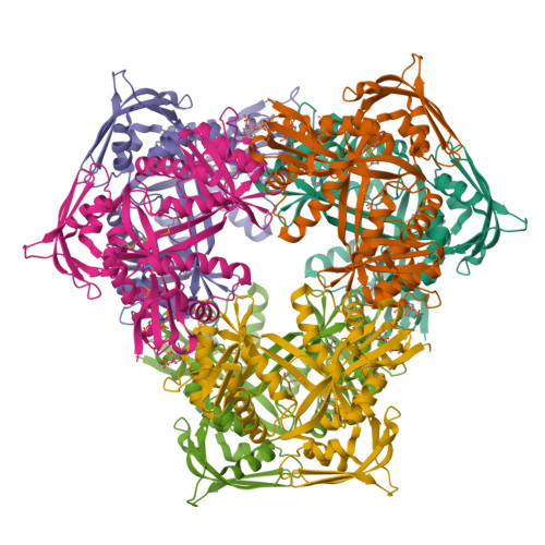
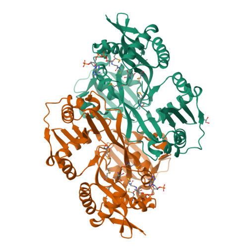
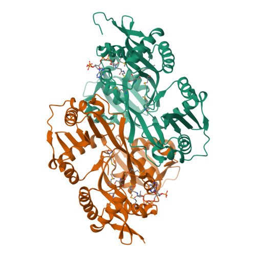
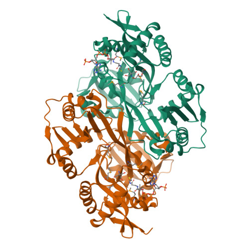
| GCN5-related N-acetyltransferase | 392 | Kribbella flavida DSM 17836 | Mutation(s): 0 Gene Names: Kfla_4406 | 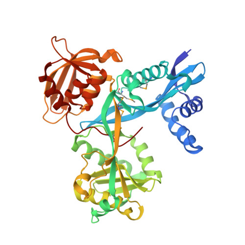 | |
UniProt | |||||
Find proteins for D2PVF8 (Kribbella flavida (strain DSM 17836 / JCM 10339 / NBRC 14399)) Explore D2PVF8 Go to UniProtKB: D2PVF8 | |||||
Entity Groups | |||||
| Sequence Clusters | 30% Identity50% Identity70% Identity90% Identity95% Identity100% Identity | ||||
| UniProt Group | D2PVF8 | ||||
Sequence AnnotationsExpand | |||||
| |||||
| Ligands 4 Unique | |||||
|---|---|---|---|---|---|
| ID | Chains | Name / Formula / InChI Key | 2D Diagram | 3D Interactions | |
| AC8 Query on AC8 | L [auth C] | [(2R,3R,4R,5R)-5-(6-amino-9H-purin-9-yl)-3,4-bis(phosphonooxy)tetrahydrofuran-2-yl]methyl (3R)-3-hydroxy-2,2-dimethyl-4-oxo-4-({3-oxo-3-[(2-sulfanylethyl)amino]propyl}amino)butyl dihydrogen diphosphate C21 H37 N7 O19 P4 S SAXULFDJRFDTDN-IBOSZNHHSA-N |  | ||
| ACO Query on ACO | G [auth A], J [auth B], N [auth D], P [auth E], S [auth F] | ACETYL COENZYME *A C23 H38 N7 O17 P3 S ZSLZBFCDCINBPY-ZSJPKINUSA-N |  | ||
| TRS Query on TRS | H [auth A] K [auth B] M [auth C] O [auth D] Q [auth E] | 2-AMINO-2-HYDROXYMETHYL-PROPANE-1,3-DIOL C4 H12 N O3 LENZDBCJOHFCAS-UHFFFAOYSA-O |  | ||
| GOL Query on GOL | I [auth A], R [auth E] | GLYCEROL C3 H8 O3 PEDCQBHIVMGVHV-UHFFFAOYSA-N |  | ||
| Modified Residues 1 Unique | |||||
|---|---|---|---|---|---|
| ID | Chains | Type | Formula | 2D Diagram | Parent |
| MSE Query on MSE | A, B, C, D, E | L-PEPTIDE LINKING | C5 H11 N O2 Se |  | MET |
| Length ( Å ) | Angle ( ˚ ) |
|---|---|
| a = 92.138 | α = 90 |
| b = 166.518 | β = 90 |
| c = 183.613 | γ = 90 |
| Software Name | Purpose |
|---|---|
| SBC-Collect | data collection |
| HKL-3000 | data collection |
| HKL-3000 | phasing |
| SHELXD | phasing |
| MLPHARE | phasing |
| DM | model building |
| RESOLVE | model building |
| Coot | model building |
| PHENIX | refinement |
| HKL-3000 | data reduction |
| HKL-3000 | data scaling |
| DM | phasing |
| RESOLVE | phasing |
RCSB PDB is hosted by

RCSB PDB is a member of the