4O2L

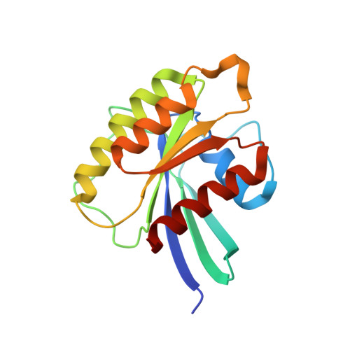
Structure of Mus musculus Rheb G63A mutant bound to GTP


Experimental Data Snapshot

  • Method: X-RAY DIFFRACTION
  • Resolution: 2.40 Å
  • R-Value Free: 0.243 
  • R-Value Work: 0.195 

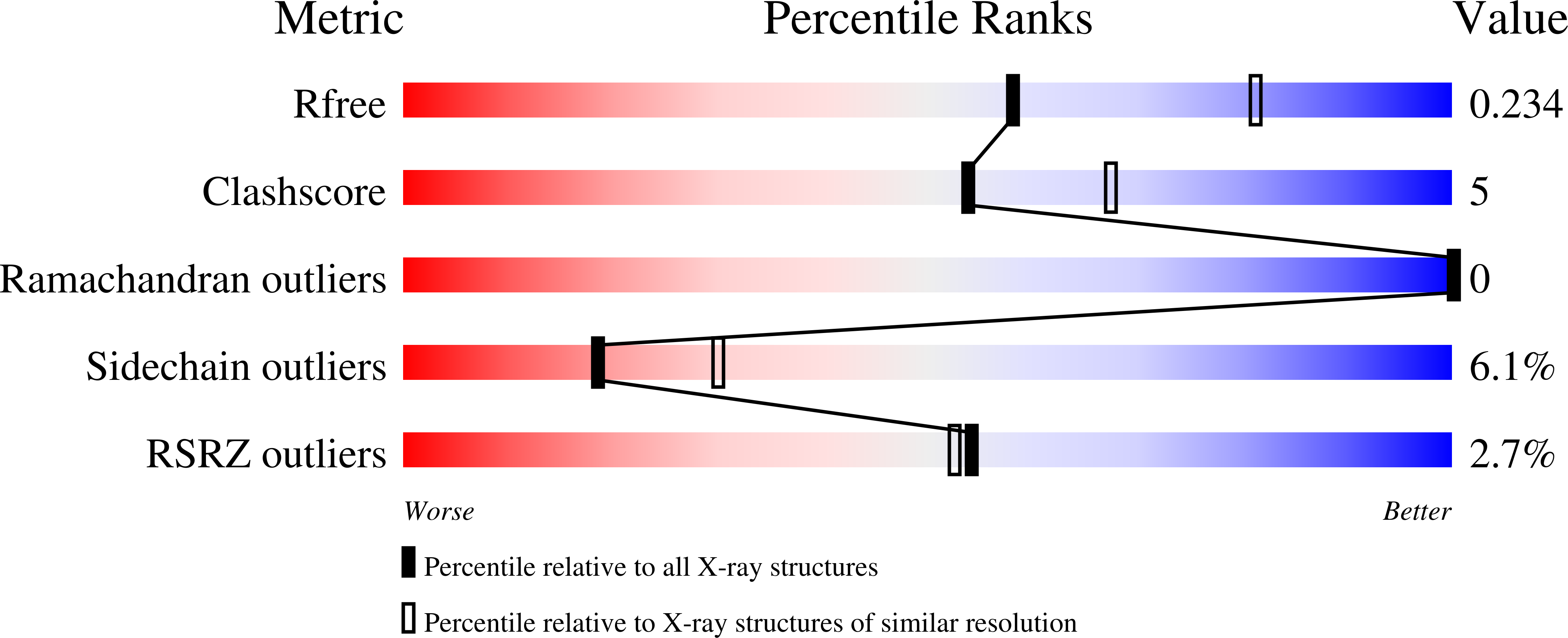
wwPDB Validation   3D Report Full Report


Ligand Structure Quality Assessment 


This is version 1.2 of the entry. See complete history


Literature

Structure-guided mutation of the conserved G3-box glycine in Rheb generates a constitutively activated regulator of mammalian target of rapamycin (mTOR).

Mazhab-Jafari, M.T.Marshall, C.B.Ho, J.Ishiyama, N.Stambolic, V.Ikura, M.

(2014) J Biol Chem 289: 12195-12201

  • DOI: https://doi.org/10.1074/jbc.C113.543736
  • Primary Citation of Related Structures:  
    4O25, 4O2L, 4O2R

  • PubMed Abstract: 

    Constitutively activated variants of small GTPases, which provide valuable functional probes of their role in cellular signaling pathways, can often be generated by mutating the canonical catalytic residue (e.g. Ras Q61L) to impair GTP hydrolysis. However, this general approach is ineffective for a substantial fraction of the small GTPase family in which this residue is not conserved (e.g. Rap) or not catalytic (e.g. Rheb). Using a novel engineering approach, we have manipulated nucleotide binding through structure-guided substitutions of an ultraconserved glycine residue in the G3-box motif (DXXG). Substitution of Rheb Gly-63 with alanine impaired both intrinsic and TSC2 GTPase-activating protein (GAP)-mediated GTP hydrolysis by displacing the hydrolytic water molecule, whereas introduction of a bulkier valine side chain selectively blocked GTP binding by steric occlusion of the γ-phosphate. Rheb G63A stimulated phosphorylation of the mTORC1 substrate p70S6 kinase more strongly than wild-type, thus offering a new tool for mammalian target of rapamycin (mTOR) signaling.


  • Organizational Affiliation

    From the Campbell Family Cancer Research Institute, Princess Margaret Cancer Centre and, Department of Medical Biophysics, University of Toronto, Toronto, Ontario M5G 2M9, Canada.


Macromolecules
Find similar proteins by:  (by identity cutoff)  |  3D Structure
Entity ID: 1
MoleculeChains Sequence LengthOrganismDetailsImage
GTP-binding protein Rheb
A, B
171Mus musculusMutation(s): 1 
Gene Names: Rheb
EC: 3.6.5
UniProt
Find proteins for Q921J2 (Mus musculus)
Explore Q921J2 
Go to UniProtKB:  Q921J2
Entity Groups  
Sequence Clusters30% Identity50% Identity70% Identity90% Identity95% Identity100% Identity
UniProt GroupQ921J2
Sequence Annotations
Expand
  • Reference Sequence
Small Molecules
Ligands 4 Unique
IDChains Name / Formula / InChI Key2D Diagram3D Interactions
GTP
Query on GTP

Download Ideal Coordinates CCD File 
C [auth A],
E [auth B]
GUANOSINE-5'-TRIPHOSPHATE
C10 H16 N5 O14 P3
XKMLYUALXHKNFT-UUOKFMHZSA-N
TRS
Query on TRS

Download Ideal Coordinates CCD File 
G [auth B]2-AMINO-2-HYDROXYMETHYL-PROPANE-1,3-DIOL
C4 H12 N O3
LENZDBCJOHFCAS-UHFFFAOYSA-O
ACT
Query on ACT

Download Ideal Coordinates CCD File 
H [auth B]ACETATE ION
C2 H3 O2
QTBSBXVTEAMEQO-UHFFFAOYSA-M
MG
Query on MG

Download Ideal Coordinates CCD File 
D [auth A],
F [auth B]
MAGNESIUM ION
Mg
JLVVSXFLKOJNIY-UHFFFAOYSA-N
Experimental Data & Validation

Experimental Data

  • Method: X-RAY DIFFRACTION
  • Resolution: 2.40 Å
  • R-Value Free: 0.243 
  • R-Value Work: 0.195 
  • Space Group: P 2 21 21
Unit Cell:
Length ( Å )Angle ( ˚ )
a = 57.557α = 90
b = 70.248β = 90
c = 79.733γ = 90
Software Package:
Software NamePurpose
PHENIXmodel building
PHENIXrefinement
HKL-2000data reduction
HKL-2000data scaling
PHENIXphasing

Structure Validation

View Full Validation Report


Ligand Structure Quality Assessment 


Entry History 

Deposition Data

Revision History  (Full details and data files)

  • Version 1.0: 2014-03-26
    Type: Initial release
  • Version 1.1: 2014-12-24
    Changes: Database references
  • Version 1.2: 2024-02-28
    Changes: Data collection, Database references, Derived calculations