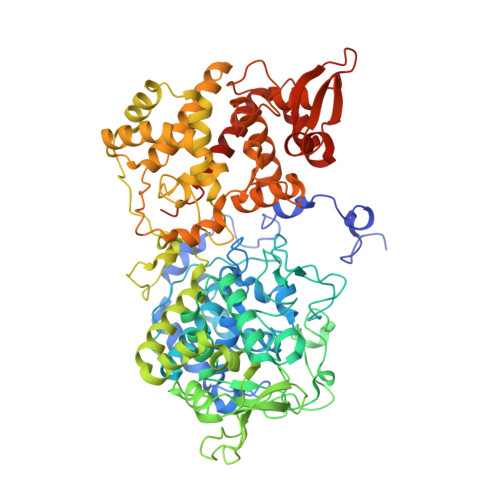
4PAE

Crystal structure of catalase-peroxidase (KatG) W78F mutant from Synechococcus elongatus PCC7942


Experimental Data Snapshot

  • Method: X-RAY DIFFRACTION
  • Resolution: 3.21 Å
  • R-Value Free: 0.260 
  • R-Value Work: 0.180 
  • R-Value Observed: 0.184 

wwPDB Validation   3D Report Full Report


Ligand Structure Quality Assessment 


This is version 1.2 of the entry. See complete history


Literature

Crystal structure of the catalase-peroxidase KatG W78F mutant from Synechococcus elongatus PCC7942 in complex with the antitubercular pro-drug isoniazid.

Kamachi, S.Hirabayashi, K.Tamoi, M.Shigeoka, S.Tada, T.Wada, K.

(2015) FEBS Lett 589: 131-137

  • DOI: https://doi.org/10.1016/j.febslet.2014.11.037
  • Primary Citation of Related Structures:  
    3X16, 4PAE

  • PubMed Abstract: 

    Isoniazid (INH) is a pro-drug that has been extensively used to treat tuberculosis. INH is activated by the heme enzyme catalase-peroxidase (KatG), but the mechanism of the activation is poorly understood, in part because the INH binding site has not been clearly established. Here, we observed that a single-residue mutation of KatG from Synechococcus elongatus PCC7942 (SeKatG), W78F, enhances INH activation. The crystal structure of INH-bound KatG-W78F revealed that INH binds to the heme pocket. The results of a thermal-shift assay implied that the flexibility of the SeKatG molecule is increased by the W78F mutation, allowing the INH molecule to easily invade the heme pocket through the access channel on the γ-edge side of the heme.


  • Organizational Affiliation

    Graduate School of Science, Osaka Prefecture University, Sakai, Osaka 599-8531, Japan.


Macromolecules
Find similar proteins by:  (by identity cutoff)  |  3D Structure
Entity ID: 1
MoleculeChains Sequence LengthOrganismDetailsImage
Catalase-peroxidase720Synechococcus elongatus PCC 7942 = FACHB-805Mutation(s): 1 
Gene Names: katGSynpcc7942_1656
EC: 1.11.1.21
UniProt
Find proteins for Q31MN3 (Synechococcus elongatus (strain ATCC 33912 / PCC 7942 / FACHB-805))
Explore Q31MN3 
Go to UniProtKB:  Q31MN3
Entity Groups  
Sequence Clusters30% Identity50% Identity70% Identity90% Identity95% Identity100% Identity
UniProt GroupQ31MN3
Sequence Annotations
Expand
  • Reference Sequence
Experimental Data & Validation

Experimental Data

  • Method: X-RAY DIFFRACTION
  • Resolution: 3.21 Å
  • R-Value Free: 0.260 
  • R-Value Work: 0.180 
  • R-Value Observed: 0.184 
  • Space Group: P 41 21 2
Unit Cell:
Length ( Å )Angle ( ˚ )
a = 107.673α = 90
b = 107.673β = 90
c = 204.788γ = 90
Software Package:
Software NamePurpose
REFMACrefinement

Structure Validation

View Full Validation Report


Ligand Structure Quality Assessment 


Entry History 

Deposition Data

Revision History  (Full details and data files)

  • Version 1.0: 2015-01-14
    Type: Initial release
  • Version 1.1: 2017-11-01
    Changes: Author supporting evidence, Data collection, Database references, Derived calculations, Other, Source and taxonomy
  • Version 1.2: 2023-12-27
    Changes: Data collection, Database references, Refinement description