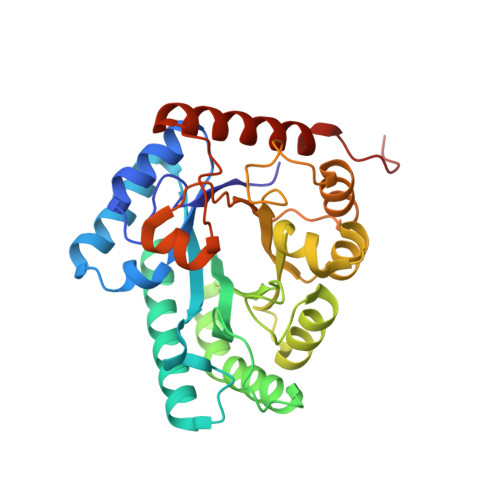
4PFH

Crystal structure of engineered D-tagatose 3-epimerase PcDTE-IDF8


Experimental Data Snapshot

  • Method: X-RAY DIFFRACTION
  • Resolution: 1.90 Å
  • R-Value Free: 0.177 
  • R-Value Work: 0.144 
  • R-Value Observed: 0.146 

wwPDB Validation   3D Report Full Report


Ligand Structure Quality Assessment 


This is version 1.5 of the entry. See complete history


Literature

Directed Divergent Evolution of a Thermostable D-Tagatose Epimerase towards Improved Activity for Two Hexose Substrates.

Bosshart, A.Hee, C.S.Bechtold, M.Schirmer, T.Panke, S.

(2015) Chembiochem 16: 592-601

  • DOI: https://doi.org/10.1002/cbic.201402620
  • Primary Citation of Related Structures:  
    4PFH, 4PGL, 4Q7I

  • PubMed Abstract: 

    Functional promiscuity of enzymes can often be harnessed as the starting point for the directed evolution of novel biocatalysts. Here we describe the divergent morphing of an engineered thermostable variant (Var8) of a promiscuous D-tagatose epimerase (DTE) into two efficient catalysts for the C3 epimerization of D-fructose to D-psicose and of L-sorbose to L-tagatose. Iterative single-site randomization and screening of 48 residues in the first and second shells around the substrate-binding site of Var8 yielded the eight-site mutant IDF8 (ninefold improved kcat for the epimerization of D-fructose) and the six-site mutant ILS6 (14-fold improved epimerization of L-sorbose), compared to Var8. Structure analysis of IDF8 revealed a charged patch at the entrance of its active site; this presumably facilitates entry of the polar substrate. The improvement in catalytic activity of variant ILS6 is thought to relate to subtle changes in the hydration of the bound substrate. The structures can now be used to select additional sites for further directed evolution of the ketohexose epimerase.


  • Organizational Affiliation

    Department of Biosystems Science and Engineering, ETH Zürich, Mattenstrasse 26, 4058 Basel (Switzerland).


Macromolecules
Find similar proteins by:  (by identity cutoff)  |  3D Structure
Entity ID: 1
MoleculeChains Sequence LengthOrganismDetailsImage
D-tagatose 3-epimerase
A, B
298Pseudomonas cichoriiMutation(s): 15 
EC: 5.3.1 (PDB Primary Data), 5.1.3.31 (UniProt)
UniProt
Find proteins for O50580 (Pseudomonas cichorii)
Explore O50580 
Go to UniProtKB:  O50580
Entity Groups  
Sequence Clusters30% Identity50% Identity70% Identity90% Identity95% Identity100% Identity
UniProt GroupO50580
Sequence Annotations
Expand
  • Reference Sequence
Small Molecules
Ligands 3 Unique
IDChains Name / Formula / InChI Key2D Diagram3D Interactions
PSJ
Query on PSJ

Download Ideal Coordinates CCD File 
E [auth A],
H [auth B]
D-psicose
C6 H12 O6
BJHIKXHVCXFQLS-PUFIMZNGSA-N
FUD
Query on FUD

Download Ideal Coordinates CCD File 
F [auth A],
I [auth B]
D-fructose
C6 H12 O6
BJHIKXHVCXFQLS-UYFOZJQFSA-N
MN
Query on MN

Download Ideal Coordinates CCD File 
C [auth A],
D [auth A],
G [auth B]
MANGANESE (II) ION
Mn
WAEMQWOKJMHJLA-UHFFFAOYSA-N
Experimental Data & Validation

Experimental Data

  • Method: X-RAY DIFFRACTION
  • Resolution: 1.90 Å
  • R-Value Free: 0.177 
  • R-Value Work: 0.144 
  • R-Value Observed: 0.146 
  • Space Group: P 1 21 1
Unit Cell:
Length ( Å )Angle ( ˚ )
a = 57.46α = 90
b = 86.74β = 99.1
c = 61.82γ = 90
Software Package:
Software NamePurpose
REFMACrefinement
MOSFLMdata reduction
Aimlessdata scaling
MOLREPphasing
Cootmodel building

Structure Validation

View Full Validation Report


Ligand Structure Quality Assessment 


Entry History 

Deposition Data

Revision History  (Full details and data files)

  • Version 1.0: 2014-10-22
    Type: Initial release
  • Version 1.1: 2015-02-11
    Changes: Other
  • Version 1.2: 2015-02-18
    Changes: Database references
  • Version 1.3: 2015-03-04
    Changes: Database references
  • Version 1.4: 2017-11-22
    Changes: Derived calculations, Refinement description, Source and taxonomy, Structure summary
  • Version 1.5: 2023-12-27
    Changes: Data collection, Database references, Derived calculations, Refinement description