4PY4 | pdb_00004py4

Crystal structure of human poly(ADP-ribose) polymerase 14, catalytic domain in complex with an inhibitor XL2


Experimental Data Snapshot

  • Method: X-RAY DIFFRACTION
  • Resolution: 2.76 Å
  • R-Value Free: 
    0.257 (Depositor), 0.250 (DCC) 
  • R-Value Work: 
    0.202 (Depositor), 0.200 (DCC) 
  • R-Value Observed: 
    0.203 (Depositor) 

Starting Model: experimental
View more details

wwPDB Validation   3D Report Full Report


Ligand Structure Quality Assessment 

Created with Raphaël 2.3.0Worse 01 BetterLigand structure goodness of fit to experimental dataBest fitted XL2Click on this verticalbar to view details

This is version 1.3 of the entry. See complete history


Literature

Palladium-catalyzed N-arylation of 2-aminobenzothiazole-4-carboxylates/carboxamides: facile synthesis of PARP14 inhibitors

Wang, P.Li, J.Jiang, X.Liu, Z.Ye, N.Xu, Y.Yang, G.Xu, Y.Zhang, A.

(2017) Tetrahedron 70: 5666-5673


Macromolecules
Find similar proteins by:  (by identity cutoff)  |  3D Structure
Entity ID: 1
MoleculeChains Sequence LengthOrganismDetailsImage
Poly [ADP-ribose] polymerase 14
A, B
189Homo sapiensMutation(s): 0 
Gene Names: PARP14BAL2KIAA1268
EC: 2.4.2.30 (PDB Primary Data), 2.4.2 (UniProt)
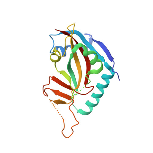
UniProt & NIH Common Fund Data Resources
Find proteins for Q460N5 (Homo sapiens)
Explore Q460N5 
Go to UniProtKB:  Q460N5
PHAROS:  Q460N5
GTEx:  ENSG00000173193 
Entity Groups  
Sequence Clusters30% Identity50% Identity70% Identity90% Identity95% Identity100% Identity
UniProt GroupQ460N5
Sequence Annotations
Expand
  • Reference Sequence
Small Molecules
Ligands 1 Unique
IDChains Name / Formula / InChI Key2D Diagram3D Interactions
XL2
Query on XL2

Download Ideal Coordinates CCD File 
C [auth A],
D [auth B]
2-({4-[(1R)-1-(dimethylamino)ethyl]phenyl}amino)-6-fluoro-1,3-benzothiazole-4-carboxamide
C18 H19 F N4 O S
ZZNKSJOSXHEVOQ-SNVBAGLBSA-N
Binding Affinity Annotations 
IDSourceBinding Affinity
XL2 BindingDB:  4PY4 IC50: 1690 (nM) from 1 assay(s)
Experimental Data & Validation

Experimental Data

  • Method: X-RAY DIFFRACTION
  • Resolution: 2.76 Å
  • R-Value Free:  0.257 (Depositor), 0.250 (DCC) 
  • R-Value Work:  0.202 (Depositor), 0.200 (DCC) 
  • R-Value Observed: 0.203 (Depositor) 
Space Group: P 21 21 21
Unit Cell:
Length ( Å )Angle ( ˚ )
a = 44.58α = 90
b = 66.91β = 90
c = 144.68γ = 90
Software Package:
Software NamePurpose
XDSdata scaling
PHENIXmodel building
PHENIXrefinement
XDSdata reduction
PHENIXphasing

Structure Validation

View Full Validation Report


Ligand Structure Quality Assessment 

Created with Raphaël 2.3.0Worse 01 BetterLigand structure goodness of fit to experimental dataBest fitted XL2Click on this verticalbar to view details

Entry History 

Deposition Data

  • Released Date: 2015-04-01 
  • Deposition Author(s): Li, J., Xu, Y.

Revision History  (Full details and data files)

  • Version 1.0: 2015-04-01
    Type: Initial release
  • Version 1.1: 2017-12-06
    Changes: Database references
  • Version 1.2: 2023-11-08
    Changes: Data collection, Database references, Derived calculations, Refinement description
  • Version 1.3: 2024-11-20
    Changes: Structure summary