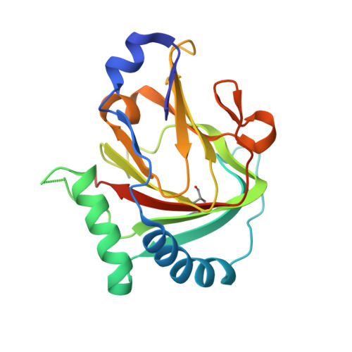
4QKD

Crystal structure of human ALKBH7 in complex with alpha-ketoglutarate and Mn(II)


Experimental Data Snapshot

  • Method: X-RAY DIFFRACTION
  • Resolution: 1.35 Å
  • R-Value Free: 0.163 
  • R-Value Work: 0.146 
  • R-Value Observed: 0.146 

Starting Model: experimental
View more details

wwPDB Validation   3D Report Full Report


This is version 1.4 of the entry. See complete history


Literature

The atomic resolution structure of human AlkB homolog 7 (ALKBH7), a key protein for programmed necrosis and fat metabolism

Wang, G.He, Q.Feng, C.Liu, Y.Deng, Z.Qi, X.Wu, W.Mei, P.Chen, Z.

(2014) J Biol Chem 289: 27924-27936

  • DOI: https://doi.org/10.1074/jbc.M114.590505
  • Primary Citation of Related Structures:  
    4QKB, 4QKD, 4QKF

  • PubMed Abstract: 

    ALKBH7 is the mitochondrial AlkB family member that is required for alkylation- and oxidation-induced programmed necrosis. In contrast to the protective role of other AlkB family members after suffering alkylation-induced DNA damage, ALKBH7 triggers the collapse of mitochondrial membrane potential and promotes cell death. Moreover, genetic ablation of mouse Alkbh7 dramatically increases body weight and fat mass. Here, we present crystal structures of human ALKBH7 in complex with Mn(II) and α-ketoglutarate at 1.35 Å or N-oxalylglycine at 2.0 Å resolution. ALKBH7 possesses the conserved double-stranded β-helix fold that coordinates a catalytically active iron by a conserved HX(D/E) … Xn … H motif. Self-hydroxylation of Leu-110 was observed, indicating that ALKBH7 has the potential to catalyze hydroxylation of its substrate. Unlike other AlkB family members whose substrates are DNA or RNA, ALKBH7 is devoid of the "nucleotide recognition lid" which is essential for binding nucleobases, and thus exhibits a solvent-exposed active site; two loops between β-strands β6 and β7 and between β9 and β10 create a special outer wall of the minor β-sheet of the double-stranded β-helix and form a negatively charged groove. These distinct features suggest that ALKBH7 may act on protein substrate rather than nucleic acids. Taken together, our findings provide a structural basis for understanding the distinct function of ALKBH7 in the AlkB family and offer a foundation for drug design in treating cell death-related diseases and metabolic diseases.


  • Organizational Affiliation

    From the State Key Laboratory of Agrobiotechnology, China Agricultural University, Beijing 100193 and.


Macromolecules
Find similar proteins by:  (by identity cutoff)  |  3D Structure
Entity ID: 1
MoleculeChains Sequence LengthOrganismDetailsImage
Alpha-ketoglutarate-dependent dioxygenase alkB homolog 7, mitochondrial
A, B, C
200Homo sapiensMutation(s): 1 
Gene Names: ABH7ALKBH7SPATA11UNQ6002/PRO34564
EC: 1.14.11
UniProt & NIH Common Fund Data Resources
Find proteins for Q9BT30 (Homo sapiens)
Explore Q9BT30 
Go to UniProtKB:  Q9BT30
PHAROS:  Q9BT30
GTEx:  ENSG00000125652 
Entity Groups  
Sequence Clusters30% Identity50% Identity70% Identity90% Identity95% Identity100% Identity
UniProt GroupQ9BT30
Sequence Annotations
Expand
  • Reference Sequence
Small Molecules
Ligands 2 Unique
IDChains Name / Formula / InChI Key2D Diagram3D Interactions
AKG
Query on AKG

Download Ideal Coordinates CCD File 
E [auth A],
H [auth B],
L [auth C]
2-OXOGLUTARIC ACID
C5 H6 O5
KPGXRSRHYNQIFN-UHFFFAOYSA-N
MN
Query on MN

Download Ideal Coordinates CCD File 
D [auth A]
F [auth A]
G [auth B]
I [auth B]
J [auth B]
D [auth A],
F [auth A],
G [auth B],
I [auth B],
J [auth B],
K [auth C],
M [auth C]
MANGANESE (II) ION
Mn
WAEMQWOKJMHJLA-UHFFFAOYSA-N
Modified Residues  1 Unique
IDChains TypeFormula2D DiagramParent
LED
Query on LED
A, B, C
L-PEPTIDE LINKINGC6 H11 N O3LEU
Experimental Data & Validation

Experimental Data

  • Method: X-RAY DIFFRACTION
  • Resolution: 1.35 Å
  • R-Value Free: 0.163 
  • R-Value Work: 0.146 
  • R-Value Observed: 0.146 
  • Space Group: P 1 21 1
Unit Cell:
Length ( Å )Angle ( ˚ )
a = 66.426α = 90
b = 82.048β = 120.03
c = 66.414γ = 90
Software Package:
Software NamePurpose
ADSCdata collection
MOLREPphasing
REFMACrefinement
HKL-2000data reduction
HKL-2000data scaling

Structure Validation

View Full Validation Report


Entry History 

Deposition Data

Revision History  (Full details and data files)

  • Version 1.0: 2014-08-20
    Type: Initial release
  • Version 1.1: 2015-10-21
    Changes: Database references
  • Version 1.2: 2023-11-08
    Changes: Data collection, Database references, Derived calculations, Refinement description
  • Version 1.3: 2023-12-06
    Changes: Data collection
  • Version 1.4: 2024-10-30
    Changes: Structure summary