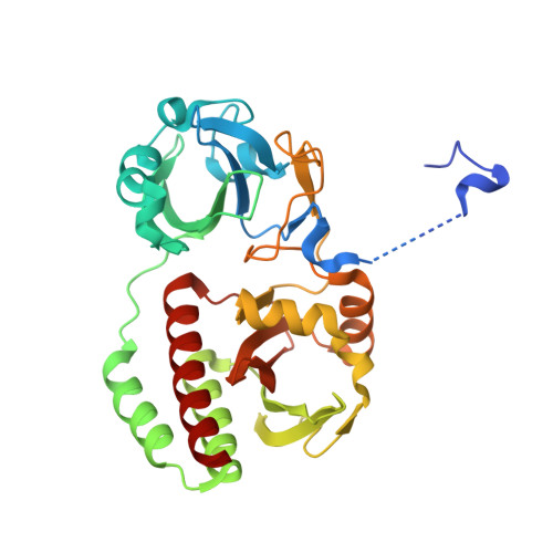
4XTQ

Crystal structure of a mutant (C20S) of a near-infrared fluorescent protein BphP1-FP


Experimental Data Snapshot

  • Method: X-RAY DIFFRACTION
  • Resolution: 1.64 Å
  • R-Value Free: 0.208 
  • R-Value Work: 0.175 
  • R-Value Observed: 0.177 

wwPDB Validation   3D Report Full Report


Ligand Structure Quality Assessment 


This is version 2.0 of the entry. See complete history


Literature

Molecular Basis of Spectral Diversity in Near-Infrared Phytochrome-Based Fluorescent Proteins.

Shcherbakova, D.M.Baloban, M.Pletnev, S.Malashkevich, V.N.Xiao, H.Dauter, Z.Verkhusha, V.V.

(2015) Chem Biol 22: 1540-1551

  • DOI: https://doi.org/10.1016/j.chembiol.2015.10.007
  • Primary Citation of Related Structures:  
    4XTQ

  • PubMed Abstract: 

    Near-infrared fluorescent proteins (NIR FPs) engineered from bacterial phytochromes (BphPs) are the probes of choice for deep-tissue imaging. Detection of several processes requires spectrally distinct NIR FPs. We developed an NIR FP, BphP1-FP, which has the most blue-shifted spectra and the highest fluorescence quantum yield among BphP-derived FPs. We found that these properties result from the binding of the biliverdin chromophore to a cysteine residue in the GAF domain, unlike natural BphPs and other BphP-based FPs. To elucidate the molecular basis of the spectral shift, we applied biochemical, structural and mass spectrometry analyses and revealed the formation of unique chromophore species. Mutagenesis of NIR FPs of different origins indicated that the mechanism of the spectral shift is general and can be used to design multicolor NIR FPs from other BphPs. We applied pairs of spectrally distinct point cysteine mutants to multicolor cell labeling and demonstrated that they perform well in model deep-tissue imaging.


  • Organizational Affiliation

    Department of Anatomy and Structural Biology, Albert Einstein College of Medicine, Bronx, NY 10461, USA.


Macromolecules
Find similar proteins by:  (by identity cutoff)  |  3D Structure
Entity ID: 1
MoleculeChains Sequence LengthOrganismDetailsImage
BphP1-FP/C20S327Rhodopseudomonas palustrisMutation(s): 0 
Entity Groups  
Sequence Clusters30% Identity50% Identity70% Identity90% Identity95% Identity100% Identity
Sequence Annotations
Expand
  • Reference Sequence
Experimental Data & Validation

Experimental Data

  • Method: X-RAY DIFFRACTION
  • Resolution: 1.64 Å
  • R-Value Free: 0.208 
  • R-Value Work: 0.175 
  • R-Value Observed: 0.177 
  • Space Group: P 21 21 21
Unit Cell:
Length ( Å )Angle ( ˚ )
a = 52.656α = 90
b = 53.113β = 90
c = 107.07γ = 90
Software Package:
Software NamePurpose
REFMACrefinement
DENZOdata reduction
PDB_EXTRACTdata extraction
SCALEPACKdata scaling
MOLREPphasing

Structure Validation

View Full Validation Report


Ligand Structure Quality Assessment 


Entry History & Funding Information

Deposition Data


Funding OrganizationLocationGrant Number
National Institutes of Health/National Cancer Institute (NIH/NCI)United StatesCA164468
National Institutes of Health/National Institute of General Medical Sciences (NIH/NIGMS)United StatesGM073913
National Institutes of Health/National Institute of General Medical Sciences (NIH/NIGMS)United StatesGM108579
EU FP7 ProgramFinlandERC-2013-ADG-340233
National Institutes of Health/National Cancer Institute (NIH/NCI)United StatesHHSN261200800001E

Revision History  (Full details and data files)

  • Version 1.0: 2015-12-09
    Type: Initial release
  • Version 1.1: 2015-12-16
    Changes: Database references
  • Version 1.2: 2017-09-06
    Changes: Author supporting evidence, Database references, Derived calculations
  • Version 1.3: 2019-12-04
    Changes: Author supporting evidence
  • Version 2.0: 2024-12-25
    Changes: Advisory, Atomic model, Data collection, Database references, Derived calculations, Structure summary