4YQE

Crystal structure of E. coli WrbA in complex with benzoquinone


Experimental Data Snapshot

  • Method: X-RAY DIFFRACTION
  • Resolution: 1.33 Å
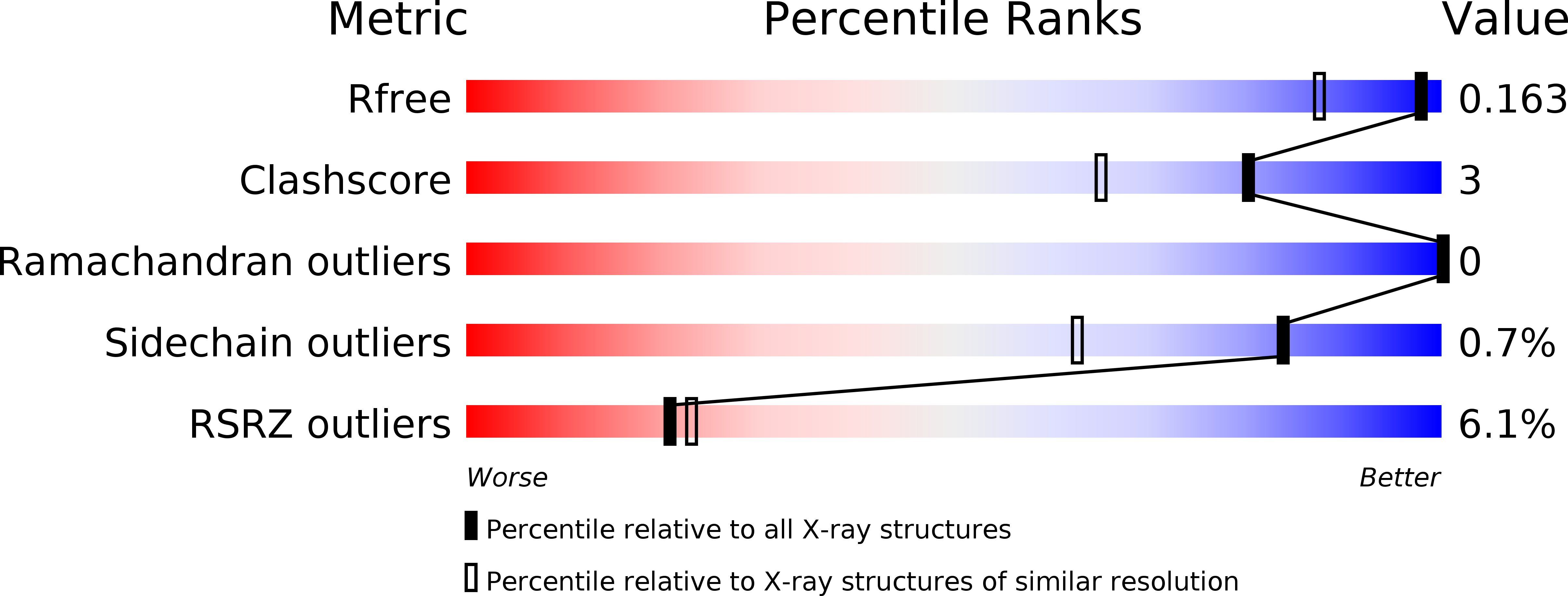
  • R-Value Free: 0.161 
  • R-Value Work: 0.125 
  • R-Value Observed: 0.127 

Starting Model: experimental
View more details

wwPDB Validation   3D Report Full Report


Ligand Structure Quality Assessment 


This is version 1.6 of the entry. See complete history


Literature

Quantum Calculations Indicate Effective Electron Transfer between FMN and Benzoquinone in a New Crystal Structure of Escherichia coli WrbA.

Degtjarik, O.Brynda, J.Ettrichova, O.Kuty, M.Sinha, D.Kuta Smatanova, I.Carey, J.Ettrich, R.Reha, D.

(2016) J Phys Chem B 120: 4867-4877

  • DOI: https://doi.org/10.1021/acs.jpcb.5b11958
  • Primary Citation of Related Structures:  
    4YQE, 5F12

  • PubMed Abstract: 

    Quantum mechanical calculations using the Marcus equation are applied to compare the electron-transfer probability for two distinct crystal structures of the Escherichia coli protein WrbA, an FMN-dependent quinone oxidoreductase, with the bound substrate benzoquinone. The calculations indicate that the position of benzoquinone in a new structure reported here and solved at 1.33 Å resolution is more likely to be relevant for the physiological reaction of WrbA than a previously reported crystal structure in which benzoquinone is shifted by ∼5 Å. Because the true electron-acceptor substrate for WrbA is not yet known, the present results can serve to constrain computational docking attempts with potential substrates that may aid in identifying the natural substrate(s) and physiological role(s) of this enzyme. The approach used here highlights a role for quantum mechanical calculations in the interpretation of protein crystal structures.


  • Organizational Affiliation

    Center for Nanobiology and Structural Biology, Institute of Microbiology, Academy of Sciences of the Czech Republic , Zamek 136, CZ-373 33 Nove Hrady, Czech Republic.


Macromolecules
Find similar proteins by:  (by identity cutoff)  |  3D Structure
Entity ID: 1
MoleculeChains Sequence LengthOrganismDetailsImage
NAD(P)H dehydrogenase (quinone)
A, B
197Escherichia coli K-12Mutation(s): 0 
Gene Names: wrbAb1004JW0989
EC: 1.6.5.2
UniProt
Find proteins for P0A8G6 (Escherichia coli (strain K12))
Explore P0A8G6 
Go to UniProtKB:  P0A8G6
Entity Groups  
Sequence Clusters30% Identity50% Identity70% Identity90% Identity95% Identity100% Identity
UniProt GroupP0A8G6
Sequence Annotations
Expand
  • Reference Sequence
Small Molecules
Modified Residues  1 Unique
IDChains TypeFormula2D DiagramParent
MHO
Query on MHO
A, B
L-PEPTIDE LINKINGC5 H11 N O3 SMET
Experimental Data & Validation

Experimental Data

  • Method: X-RAY DIFFRACTION
  • Resolution: 1.33 Å
  • R-Value Free: 0.161 
  • R-Value Work: 0.125 
  • R-Value Observed: 0.127 
  • Space Group: P 41 21 2
Unit Cell:
Length ( Å )Angle ( ˚ )
a = 61.179α = 90
b = 61.179β = 90
c = 169.992γ = 90
Software Package:
Software NamePurpose
REFMACrefinement
MOLREPphasing
PDB_EXTRACTdata extraction
XDSdata reduction
Cootmodel building
XSCALEdata scaling

Structure Validation

View Full Validation Report


Ligand Structure Quality Assessment 


Entry History 

Deposition Data

Revision History  (Full details and data files)

  • Version 1.0: 2016-05-25
    Type: Initial release
  • Version 1.1: 2016-06-01
    Changes: Database references
  • Version 1.2: 2016-06-08
    Changes: Database references
  • Version 1.3: 2016-06-22
    Changes: Database references
  • Version 1.4: 2018-01-31
    Changes: Database references
  • Version 1.5: 2024-01-10
    Changes: Data collection, Database references, Refinement description
  • Version 1.6: 2024-10-23
    Changes: Structure summary