4Z1V

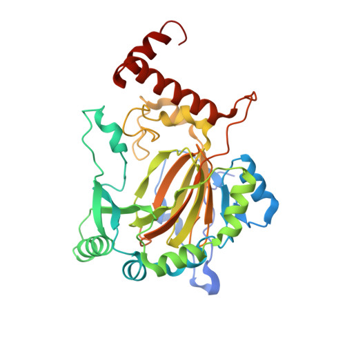
Structure of Factor Inhibiting HIF (FIH) in complex with Fe, NO, and NOG


Experimental Data Snapshot

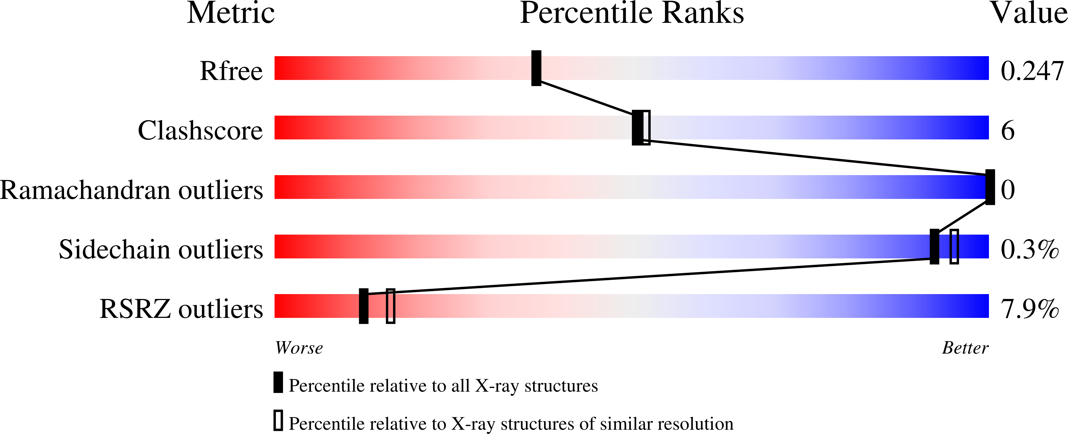
  • Method: X-RAY DIFFRACTION
  • Resolution: 2.10 Å
  • R-Value Free: 0.240 
  • R-Value Work: 0.190 
  • R-Value Observed: 0.195 

Starting Model: experimental
View more details

wwPDB Validation   3D Report Full Report


This is version 1.5 of the entry. See complete history


Literature

Substrate Promotes Productive Gas Binding in the alpha-Ketoglutarate-Dependent Oxygenase FIH.

Taabazuing, C.Y.Fermann, J.Garman, S.Knapp, M.J.

(2016) Biochemistry 55: 277-286

  • DOI: https://doi.org/10.1021/acs.biochem.5b01003
  • Primary Citation of Related Structures:  
    4Z1V, 4Z2W

  • PubMed Abstract: 

    The Fe(2+)/α-ketoglutarate (αKG)-dependent oxygenases use molecular oxygen to conduct a wide variety of reactions with important biological implications, such as DNA base excision repair, histone demethylation, and the cellular hypoxia response. These enzymes follow a sequential mechanism in which O2 binds and reacts after the primary substrate binds, making those structural factors that promote productive O2 binding central to their chemistry. A large challenge in this field is to identify strategies that engender productive turnover. Factor inhibiting HIF (FIH) is a Fe(2+)/αKG-dependent oxygenase that forms part of the O2 sensing machinery in human cells by hydroxylating the C-terminal transactivation domain (CTAD) found within the HIF-1α protein. The structure of FIH was determined with the O2 analogue NO bound to Fe, offering the first direct insight into the gas binding geometry in this enzyme. Through a combination of density functional theory calculations, {FeNO}(7) electron paramagnetic resonance spectroscopy, and ultraviolet-visible absorption spectroscopy, we demonstrate that CTAD binding stimulates O2 reactivity by altering the orientation of the bound gas molecule. Although unliganded FIH binds NO with moderate affinity, the bound gas can adopt either of two orientations with similar stability; upon CTAD binding, NO adopts a single preferred orientation that is appropriate for supporting oxidative decarboxylation. Combined with other studies of related enzymes, our data suggest that substrate-induced reorientation of bound O2 is the mechanism utilized by the αKG oxygenases to tightly couple O2 activation to substrate hydroxylation.


  • Organizational Affiliation

    Department of Chemistry, University of Massachusetts , Amherst, Massachusetts 01003, United States.


Macromolecules
Find similar proteins by:  (by identity cutoff)  |  3D Structure
Entity ID: 1
MoleculeChains Sequence LengthOrganismDetailsImage
Hypoxia-inducible factor 1-alpha inhibitor352Homo sapiensMutation(s): 0 
Gene Names: HIF1ANFIH1
EC: 1.14.11.30 (PDB Primary Data), 1.14.11 (PDB Primary Data)
UniProt & NIH Common Fund Data Resources
Find proteins for Q9NWT6 (Homo sapiens)
Explore Q9NWT6 
Go to UniProtKB:  Q9NWT6
PHAROS:  Q9NWT6
GTEx:  ENSG00000166135 
Entity Groups  
Sequence Clusters30% Identity50% Identity70% Identity90% Identity95% Identity100% Identity
UniProt GroupQ9NWT6
Sequence Annotations
Expand
  • Reference Sequence
Small Molecules
Ligands 5 Unique
IDChains Name / Formula / InChI Key2D Diagram3D Interactions
OGA
Query on OGA

Download Ideal Coordinates CCD File 
B [auth A]N-OXALYLGLYCINE
C4 H5 N O5
BIMZLRFONYSTPT-UHFFFAOYSA-N
PEG
Query on PEG

Download Ideal Coordinates CCD File 
D [auth A]DI(HYDROXYETHYL)ETHER
C4 H10 O3
MTHSVFCYNBDYFN-UHFFFAOYSA-N
SO4
Query on SO4

Download Ideal Coordinates CCD File 
F [auth A],
G [auth A],
H [auth A],
I [auth A],
J [auth A]
SULFATE ION
O4 S
QAOWNCQODCNURD-UHFFFAOYSA-L
FE
Query on FE

Download Ideal Coordinates CCD File 
E [auth A]FE (III) ION
Fe
VTLYFUHAOXGGBS-UHFFFAOYSA-N
NO
Query on NO

Download Ideal Coordinates CCD File 
C [auth A]NITRIC OXIDE
N O
ODUCDPQEXGNKDN-UHFFFAOYSA-N
Experimental Data & Validation

Experimental Data

  • Method: X-RAY DIFFRACTION
  • Resolution: 2.10 Å
  • R-Value Free: 0.240 
  • R-Value Work: 0.190 
  • R-Value Observed: 0.195 
  • Space Group: P 41 21 2
Unit Cell:
Length ( Å )Angle ( ˚ )
a = 86.65α = 90
b = 86.65β = 90
c = 147.73γ = 90
Software Package:
Software NamePurpose
REFMACrefinement
SCALAdata scaling
PDB_EXTRACTdata extraction
MOSFLMdata reduction
PHASERphasing

Structure Validation

View Full Validation Report


Entry History & Funding Information

Deposition Data


Funding OrganizationLocationGrant Number
National Institutes of Health/National Institute of General Medical Sciences (NIH/NIGMS)United StatesGM077413
National Institutes of Health/National Institute of General Medical Sciences (NIH/NIGMS)United States5T32GM008515-17

Revision History  (Full details and data files)

  • Version 1.0: 2016-01-13
    Type: Initial release
  • Version 1.1: 2016-01-20
    Changes: Data collection
  • Version 1.2: 2016-01-27
    Changes: Database references
  • Version 1.3: 2017-09-27
    Changes: Author supporting evidence, Database references, Derived calculations
  • Version 1.4: 2019-12-25
    Changes: Author supporting evidence
  • Version 1.5: 2023-09-27
    Changes: Data collection, Database references, Refinement description