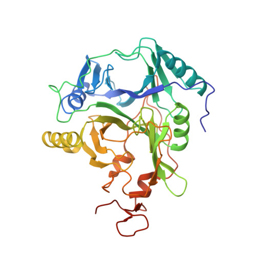
5BWG

Structure of H200C variant of Homoprotocatechuate 2,3-Dioxygenase from B.fuscum at 1.75 Ang resolution

  • Classification: OXIDOREDUCTASE
  • Organism(s): Brevibacterium fuscum
  • Expression System: Escherichia coli
  • Mutation(s): Yes 

  • Deposited: 2015-06-08 Released: 2015-10-28 
  • Deposition Author(s): Kovaleva, E.G., Lipscomb, J.D.
  • Funding Organization(s): Biotechnology and Biological Sciences Research Council (BBSRC), National Institutes of Health/National Institute of General Medical Sciences (NIH/NIGMS)

Experimental Data Snapshot

  • Method: X-RAY DIFFRACTION
  • Resolution: 1.75 Å
  • R-Value Free: 0.179 
  • R-Value Work: 0.145 
  • R-Value Observed: 0.146 

Starting Model: experimental
View more details

wwPDB Validation   3D Report Full Report


Ligand Structure Quality Assessment 


This is version 1.6 of the entry. See complete history


Literature

A Long-Lived Fe(III)-(Hydroperoxo) Intermediate in the Active H200C Variant of Homoprotocatechuate 2,3-Dioxygenase: Characterization by Mossbauer, Electron Paramagnetic Resonance, and Density Functional Theory Methods.

Meier, K.K.Rogers, M.S.Kovaleva, E.G.Mbughuni, M.M.Bominaar, E.L.Lipscomb, J.D.Munck, E.

(2015) Inorg Chem 54: 10269-10280

  • DOI: https://doi.org/10.1021/acs.inorgchem.5b01576
  • Primary Citation of Related Structures:  
    5BWG, 5BWH

  • PubMed Abstract: 

    The extradiol-cleaving dioxygenase homoprotocatechuate 2,3-dioxygenase (HPCD) binds substrate homoprotocatechuate (HPCA) and O2 sequentially in adjacent ligand sites of the active site Fe(II). Kinetic and spectroscopic studies of HPCD have elucidated catalytic roles of several active site residues, including the crucial acid-base chemistry of His200. In the present study, reaction of the His200Cys (H200C) variant with native substrate HPCA resulted in a decrease in both kcat and the rate constants for the activation steps following O2 binding by >400 fold. The reaction proceeds to form the correct extradiol product. This slow reaction allowed a long-lived (t1/2 = 1.5 min) intermediate, H200C-HPCAInt1 (Int1), to be trapped. Mössbauer and parallel mode electron paramagnetic resonance (EPR) studies show that Int1 contains an S1 = 5/2 Fe(III) center coupled to an SR = 1/2 radical to give a ground state with total spin S = 2 (J > 40 cm(-1)) in Hexch = JŜ1·ŜR. Density functional theory (DFT) property calculations for structural models suggest that Int1 is a (HPCA semiquinone(•))Fe(III)(OOH) complex, in which OOH is protonated at the distal O and the substrate hydroxyls are deprotonated. By combining Mössbauer and EPR data of Int1 with DFT calculations, the orientations of the principal axes of the (57)Fe electric field gradient and the zero-field splitting tensors (D = 1.6 cm(-1), E/D = 0.05) were determined. This information was used to predict hyperfine splittings from bound (17)OOH. DFT reactivity analysis suggests that Int1 can evolve from a ferromagnetically coupled Fe(III)-superoxo precursor by an inner-sphere proton-coupled-electron-transfer process. Our spectroscopic and DFT results suggest that a ferric hydroperoxo species is capable of extradiol catalysis.


  • Organizational Affiliation

    Department of Chemistry, Carnegie Mellon University , Pittsburgh, Pennsylvania 15213, United States.


Macromolecules
Find similar proteins by:  (by identity cutoff)  |  3D Structure
Entity ID: 1
MoleculeChains Sequence LengthOrganismDetailsImage
Homoprotocatechuate 2,3-dioxygenase
A, B, C, D
365Brevibacterium fuscumMutation(s): 1 
UniProt
Find proteins for Q45135 (Brevibacterium fuscum)
Explore Q45135 
Go to UniProtKB:  Q45135
Entity Groups  
Sequence Clusters30% Identity50% Identity70% Identity90% Identity95% Identity100% Identity
UniProt GroupQ45135
Sequence Annotations
Expand
  • Reference Sequence
Small Molecules
Ligands 5 Unique
IDChains Name / Formula / InChI Key2D Diagram3D Interactions
P6G
Query on P6G

Download Ideal Coordinates CCD File 
G [auth A],
L [auth B],
M [auth B],
R [auth C],
U [auth D]
HEXAETHYLENE GLYCOL
C12 H26 O7
IIRDTKBZINWQAW-UHFFFAOYSA-N
MPO
Query on MPO

Download Ideal Coordinates CCD File 
H [auth A],
N [auth B],
O [auth B]
3[N-MORPHOLINO]PROPANE SULFONIC ACID
C7 H15 N O4 S
DVLFYONBTKHTER-UHFFFAOYSA-N
FE2
Query on FE2

Download Ideal Coordinates CCD File 
E [auth A],
I [auth B],
P [auth C],
S [auth D]
FE (II) ION
Fe
CWYNVVGOOAEACU-UHFFFAOYSA-N
CA
Query on CA

Download Ideal Coordinates CCD File 
J [auth B]CALCIUM ION
Ca
BHPQYMZQTOCNFJ-UHFFFAOYSA-N
CL
Query on CL

Download Ideal Coordinates CCD File 
F [auth A],
K [auth B],
Q [auth C],
T [auth D]
CHLORIDE ION
Cl
VEXZGXHMUGYJMC-UHFFFAOYSA-M
Experimental Data & Validation

Experimental Data

  • Method: X-RAY DIFFRACTION
  • Resolution: 1.75 Å
  • R-Value Free: 0.179 
  • R-Value Work: 0.145 
  • R-Value Observed: 0.146 
  • Space Group: P 21 21 2
Unit Cell:
Length ( Å )Angle ( ˚ )
a = 110.748α = 90
b = 150.627β = 90
c = 96.269γ = 90
Software Package:
Software NamePurpose
SCALAdata scaling
REFMACrefinement
PDB_EXTRACTdata extraction
XDSdata reduction
MOLREPphasing

Structure Validation

View Full Validation Report


Ligand Structure Quality Assessment 


Entry History & Funding Information

Deposition Data


Funding OrganizationLocationGrant Number
Biotechnology and Biological Sciences Research Council (BBSRC)United KingdomBB/H001905/1
National Institutes of Health/National Institute of General Medical Sciences (NIH/NIGMS)United StatesGM 24689

Revision History  (Full details and data files)

  • Version 1.0: 2015-10-28
    Type: Initial release
  • Version 1.1: 2015-11-11
    Changes: Database references
  • Version 1.2: 2017-09-06
    Changes: Author supporting evidence, Database references, Derived calculations, Structure summary
  • Version 1.3: 2017-11-22
    Changes: Database references
  • Version 1.4: 2019-12-25
    Changes: Author supporting evidence
  • Version 1.5: 2020-01-01
    Changes: Author supporting evidence
  • Version 1.6: 2023-09-27
    Changes: Data collection, Database references, Derived calculations, Refinement description