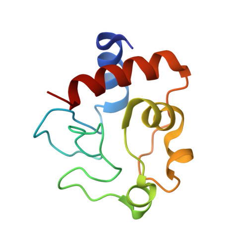
5DFS

Crystal structure of Spider Monkey Cytochrome C at 1.15 Angstrom


Experimental Data Snapshot

  • Method: X-RAY DIFFRACTION
  • Resolution: 1.15 Å
  • R-Value Free: 0.182 
  • R-Value Work: 0.160 
  • R-Value Observed: 0.160 

Starting Model: experimental
View more details

wwPDB Validation   3D Report Full Report


Ligand Structure Quality Assessment 


This is version 2.2 of the entry. See complete history


Literature

Disruption of a hydrogen bond network in human versus spider monkey cytochrome c affects heme crevice stability.

Goldes, M.E.Jeakins-Cooley, M.E.McClelland, L.J.Mou, T.C.Bowler, B.E.

(2016) J Inorg Biochem 158: 62-69

  • DOI: https://doi.org/10.1016/j.jinorgbio.2015.12.025
  • Primary Citation of Related Structures:  
    5DFS

  • PubMed Abstract: 

    The hypothesis that the recent rapid evolution of primate cytochromes c, which primarily involves residues in the least stable Ω-loop (Ω-loop C, residues 40-57), stabilizes the heme crevice of cytochrome c relative to other mammals, is tested. To accomplish this goal, we have compared the properties of human and spider monkey cytochrome c and a set of four variants produced in the process of converting human cytochrome c into spider monkey cytochrome c. The global stability of all variants has been measured by guanidine hydrochloride denaturation. The stability of the heme crevice has been assessed with the alkaline conformational transition. Structural insight into the effects of the five amino acid substitutions needed to convert human cytochrome c into spider monkey cytochrome c is provided by a 1.15Å resolution structure of spider monkey cytochrome c. The global stability for all variants is near 9.0kcal/mol at 25°C and pH7, which is higher than that observed for other mammalian cytochromes c. The heme crevice stability is more sensitive to the substitutions required to produce spider monkey cytochrome c with decreases of up to 0.5 units in the apparent pKa of the alkaline conformational transition relative to human cytochrome c. The structure of spider monkey cytochrome c indicates that the Y46F substitution destabilizes the heme crevice by disrupting an extensive hydrogen bond network that connects three surface loops including Ω-loop D (residues 70-85), which contains the Met80 heme ligand.


  • Organizational Affiliation

    Department of Chemistry and Biochemistry, University of Montana, Missoula, MT 59812, United States.


Macromolecules
Find similar proteins by:  (by identity cutoff)  |  3D Structure
Entity ID: 1
MoleculeChains Sequence LengthOrganismDetailsImage
Cytochrome c
A, B
105Ateles sp.Mutation(s): 0 
Gene Names: CYCSCYC
UniProt
Find proteins for P00003 (Ateles sp.)
Explore P00003 
Go to UniProtKB:  P00003
Entity Groups  
Sequence Clusters30% Identity50% Identity70% Identity90% Identity95% Identity100% Identity
UniProt GroupP00003
Sequence Annotations
Expand
  • Reference Sequence
Small Molecules
Ligands 3 Unique
IDChains Name / Formula / InChI Key2D Diagram3D Interactions
HEC
Query on HEC

Download Ideal Coordinates CCD File 
C [auth A],
H [auth B]
HEME C
C34 H34 Fe N4 O4
HXQIYSLZKNYNMH-LJNAALQVSA-N
EDO
Query on EDO

Download Ideal Coordinates CCD File 
D [auth A]
E [auth A]
I [auth B]
J [auth B]
K [auth B]
D [auth A],
E [auth A],
I [auth B],
J [auth B],
K [auth B],
L [auth B]
1,2-ETHANEDIOL
C2 H6 O2
LYCAIKOWRPUZTN-UHFFFAOYSA-N
CL
Query on CL

Download Ideal Coordinates CCD File 
F [auth A],
G [auth A]
CHLORIDE ION
Cl
VEXZGXHMUGYJMC-UHFFFAOYSA-M
Experimental Data & Validation

Experimental Data

  • Method: X-RAY DIFFRACTION
  • Resolution: 1.15 Å
  • R-Value Free: 0.182 
  • R-Value Work: 0.160 
  • R-Value Observed: 0.160 
  • Space Group: P 1 21 1
Unit Cell:
Length ( Å )Angle ( ˚ )
a = 31.534α = 90
b = 42.733β = 94.47
c = 67.654γ = 90
Software Package:
Software NamePurpose
PHENIXrefinement
HKL-2000data reduction
HKL-2000data scaling
PHENIXphasing

Structure Validation

View Full Validation Report


Ligand Structure Quality Assessment 


Entry History 

Deposition Data

Revision History  (Full details and data files)

  • Version 1.0: 2016-03-02
    Type: Initial release
  • Version 1.1: 2016-06-08
    Changes: Database references
  • Version 2.0: 2021-03-10
    Changes: Atomic model, Data collection, Database references, Derived calculations, Non-polymer description, Structure summary
  • Version 2.1: 2023-09-27
    Changes: Data collection, Database references, Refinement description
  • Version 2.2: 2024-10-09
    Changes: Structure summary