High-Resolution X-Ray Structures of Two Functionally Distinct Members of the Cyclic Amide Hydrolase Family of Toblerone Fold Enzymes.
Peat, T.S., Balotra, S., Wilding, M., Hartley, C.J., Newman, J., Scott, C.(2017) Appl Environ Microbiol 83
- PubMed: 28235873
- DOI: https://doi.org/10.1128/AEM.03365-16
- Primary Citation of Related Structures:
5HWE, 5HXU, 5HXZ, 5HY0, 5HY1, 5HY2, 5HY4 - PubMed Abstract:
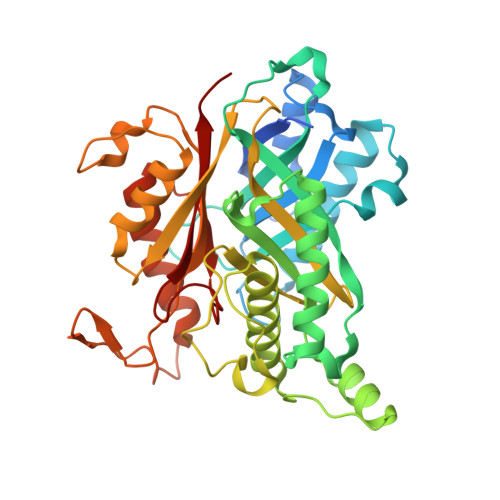
The Toblerone fold was discovered recently when the first structure of the cyclic amide hydrolase, AtzD (a cyanuric acid hydrolase), was elucidated. We surveyed the cyclic amide hydrolase family, finding a strong correlation between phylogenetic distribution and specificity for either cyanuric acid or barbituric acid. One of six classes (IV) could not be tested due to a lack of expression of the proteins from it, and another class (V) had neither cyanuric acid nor barbituric acid hydrolase activity. High-resolution X-ray structures were obtained for a class VI barbituric acid hydrolase (1.7 Å) from a Rhodococcus species and a class V cyclic amide hydrolase (2.4 Å) from a Frankia species for which we were unable to identify a substrate. Both structures were homologous with the tetrameric Toblerone fold enzyme AtzD, demonstrating a high degree of structural conservation within the cyclic amide hydrolase family. The barbituric acid hydrolase structure did not contain zinc, in contrast with early reports of zinc-dependent activity for this enzyme. Instead, each barbituric acid hydrolase monomer contained either Na + or Mg 2+ , analogous to the structural metal found in cyanuric acid hydrolase. The Frankia cyclic amide hydrolase contained no metal but instead formed unusual, reversible, intermolecular vicinal disulfide bonds that contributed to the thermal stability of the protein. The active sites were largely conserved between the three enzymes, differing at six positions, which likely determine substrate specificity. IMPORTANCE The Toblerone fold enzymes catalyze an unusual ring-opening hydrolysis with cyclic amide substrates. A survey of these enzymes shows that there is a good correlation between physiological function and phylogenetic distribution within this family of enzymes and provide insights into the evolutionary relationships between the cyanuric acid and barbituric acid hydrolases. This family of enzymes is structurally and mechanistically distinct from other enzyme families; however, to date the structure of just two, physiologically identical, enzymes from this family has been described. We present two new structures: a barbituric acid hydrolase and an enzyme of unknown function. These structures confirm that members of the CyAH family have the unusual Toblerone fold, albeit with some significant differences.
Organizational Affiliation:
CSIRO Biomedical Manufacturing, Parkville, Victoria, Australia.


