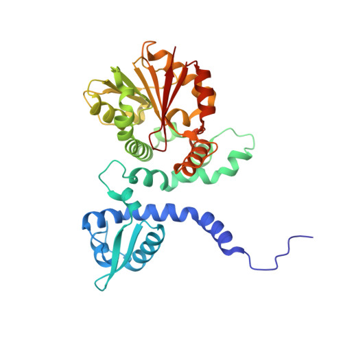
5ICF

Crystal structure of (S)-norcoclaurine 6-O-methyltransferase with S-adenosyl-L-homocysteine and sanguinarine


Experimental Data Snapshot

  • Method: X-RAY DIFFRACTION
  • Resolution: 1.80 Å
  • R-Value Free: 0.192 
  • R-Value Work: 0.156 
  • R-Value Observed: 0.158 

wwPDB Validation   3D Report Full Report


Ligand Structure Quality Assessment 


This is version 1.2 of the entry. See complete history


Literature

Crystal structure of norcoclaurine-6-O-methyltransferase, a key rate-limiting step in the synthesis of benzylisoquinoline alkaloids.

Robin, A.Y.Giustini, C.Graindorge, M.Matringe, M.Dumas, R.

(2016) Plant J 87: 641-653

  • DOI: https://doi.org/10.1111/tpj.13225
  • Primary Citation of Related Structures:  
    5ICC, 5ICE, 5ICF, 5ICG

  • PubMed Abstract: 

    Growing pharmaceutical interest in benzylisoquinoline alkaloids (BIA) coupled with their chemical complexity make metabolic engineering of microbes to create alternative platforms of production an increasingly attractive proposition. However, precise knowledge of rate-limiting enzymes and negative feedback inhibition by end-products of BIA metabolism is of paramount importance for this emerging field of synthetic biology. In this work we report the structural characterization of (S)-norcoclaurine-6-O-methyltransferase (6OMT), a key rate-limiting step enzyme involved in the synthesis of reticuline, the final intermediate to be shared between the different end-products of BIA metabolism, such as morphine, papaverine, berberine and sanguinarine. Four different crystal structures of the enzyme from Thalictrum flavum (Tf 6OMT) were solved: the apoenzyme, the complex with S-adenosyl-l-homocysteine (SAH), the complexe with SAH and the substrate and the complex with SAH and a feedback inhibitor, sanguinarine. The Tf 6OMT structural study provides a molecular understanding of its substrate specificity, active site structure and reaction mechanism. This study also clarifies the inhibition of Tf 6OMT by previously suggested feedback inhibitors. It reveals its high and time-dependent sensitivity toward sanguinarine.


  • Organizational Affiliation

    Laboratoire de Physiologie Cellulaire et Végétale, Université Grenoble Alpes, CNRS UMR5168, CEA/DRF/BIG, INRA UMR 1417, 17, Avenue des Martyrs, 38054 Grenoble, France.


Macromolecules
Find similar proteins by:  (by identity cutoff)  |  3D Structure
Entity ID: 1
MoleculeChains Sequence LengthOrganismDetailsImage
(S)-norcoclaurine 6-O-methyltransferase352Thalictrum flavum subsp. glaucumMutation(s): 0 
EC: 2.1.1.128
UniProt
Find proteins for Q5C9L7 (Thalictrum flavum subsp. glaucum)
Explore Q5C9L7 
Go to UniProtKB:  Q5C9L7
Entity Groups  
Sequence Clusters30% Identity50% Identity70% Identity90% Identity95% Identity100% Identity
UniProt GroupQ5C9L7
Sequence Annotations
Expand
  • Reference Sequence
Small Molecules
Ligands 6 Unique
IDChains Name / Formula / InChI Key2D Diagram3D Interactions
SAH
Query on SAH

Download Ideal Coordinates CCD File 
B [auth A]S-ADENOSYL-L-HOMOCYSTEINE
C14 H20 N6 O5 S
ZJUKTBDSGOFHSH-WFMPWKQPSA-N
SAU
Query on SAU

Download Ideal Coordinates CCD File 
C [auth A]13-methyl[1,3]benzodioxolo[5,6-c][1,3]dioxolo[4,5-i]phenanthridin-13-ium
C20 H14 N O4
INVGWHRKADIJHF-UHFFFAOYSA-N
DMS
Query on DMS

Download Ideal Coordinates CCD File 
T [auth A],
U [auth A]
DIMETHYL SULFOXIDE
C2 H6 O S
IAZDPXIOMUYVGZ-UHFFFAOYSA-N
EDO
Query on EDO

Download Ideal Coordinates CCD File 
G [auth A]
H [auth A]
I [auth A]
J [auth A]
K [auth A]
G [auth A],
H [auth A],
I [auth A],
J [auth A],
K [auth A],
L [auth A],
M [auth A],
N [auth A],
O [auth A],
P [auth A],
Q [auth A],
R [auth A],
S [auth A]
1,2-ETHANEDIOL
C2 H6 O2
LYCAIKOWRPUZTN-UHFFFAOYSA-N
K
Query on K

Download Ideal Coordinates CCD File 
D [auth A],
E [auth A]
POTASSIUM ION
K
NPYPAHLBTDXSSS-UHFFFAOYSA-N
NA
Query on NA

Download Ideal Coordinates CCD File 
F [auth A]SODIUM ION
Na
FKNQFGJONOIPTF-UHFFFAOYSA-N
Experimental Data & Validation

Experimental Data

  • Method: X-RAY DIFFRACTION
  • Resolution: 1.80 Å
  • R-Value Free: 0.192 
  • R-Value Work: 0.156 
  • R-Value Observed: 0.158 
  • Space Group: P 21 21 2
Unit Cell:
Length ( Å )Angle ( ˚ )
a = 73.34α = 90
b = 109.09β = 90
c = 40.95γ = 90
Software Package:
Software NamePurpose
XDSdata reduction
PHASERphasing
REFMACrefinement
PDB_EXTRACTdata extraction
XSCALEdata scaling
PHASERphasing
XSCALEdata reduction

Structure Validation

View Full Validation Report


Ligand Structure Quality Assessment 


Entry History & Funding Information

Deposition Data


Funding OrganizationLocationGrant Number
INRA (French National Institute for Agricultural Research)FranceP-BV-2

Revision History  (Full details and data files)

  • Version 1.0: 2016-06-08
    Type: Initial release
  • Version 1.1: 2016-10-12
    Changes: Database references
  • Version 1.2: 2024-05-08
    Changes: Data collection, Database references, Derived calculations