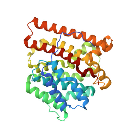
5IMI

Crystal structure of S303A Aspergillus terreus aristolochene synthase complexed with (1S,8S,9aR)-1,9a-dimethyl-8-(prop-1-en-2-yl)decahydroquinolizin-5-ium


Experimental Data Snapshot

  • Method: X-RAY DIFFRACTION
  • Resolution: 2.46 Å
  • R-Value Free: 0.251 
  • R-Value Work: 0.212 
  • R-Value Observed: 0.213 

Starting Model: experimental
View more details

wwPDB Validation   3D Report Full Report


Ligand Structure Quality Assessment 


This is version 1.4 of the entry. See complete history


Literature

Probing the Role of Active Site Water in the Sesquiterpene Cyclization Reaction Catalyzed by Aristolochene Synthase.

Chen, M.Chou, W.K.Al-Lami, N.Faraldos, J.A.Allemann, R.K.Cane, D.E.Christianson, D.W.

(2016) Biochemistry 55: 2864-2874

  • DOI: https://doi.org/10.1021/acs.biochem.6b00343
  • Primary Citation of Related Structures:  
    5IMI, 5IMN, 5IMP, 5IN8, 5IVG

  • PubMed Abstract: 

    Aristolochene synthase (ATAS) is a high-fidelity terpenoid cyclase that converts farnesyl diphosphate exclusively into the bicyclic hydrocarbon aristolochene. Previously determined crystal structures of ATAS complexes revealed trapped active site water molecules that could potentially interact with catalytic intermediates: water "w" hydrogen bonds with S303 and N299, water molecules "w1" and "w2" hydrogen bond with Q151, and a fourth water molecule coordinates to the Mg(2+)C ion. There is no obvious role for water in the ATAS mechanism because the enzyme exclusively generates a hydrocarbon product. Thus, these water molecules are tightly controlled so that they cannot react with carbocation intermediates. Steady-state kinetics and product distribution analyses of eight ATAS mutants designed to perturb interactions with active site water molecules (S303A, S303H, S303D, N299A, N299L, N299A/S303A, Q151H, and Q151E) indicate relatively modest effects on catalysis but significant effects on sesquiterpene product distributions. X-ray crystal structures of S303A, N299A, N299A/S303A, and Q151H mutants reveal minimal perturbation of active site solvent structure. Seven of the eight mutants generate farnesol and nerolidol, possibly resulting from addition of the Mg(2+)C-bound water molecule to the initially formed farnesyl cation, but no products are generated that would suggest enhanced reactivity of other active site water molecules. However, intermediate germacrene A tends to accumulate in these mutants. Thus, apart from the possible reactivity of Mg(2+)C-bound water, active site water molecules in ATAS are not directly involved in the chemistry of catalysis but instead contribute to the template that governs the conformation of the flexible substrate and carbocation intermediates.


  • Organizational Affiliation

    Roy and Diana Vagelos Laboratories, Department of Chemistry, University of Pennsylvania , Philadelphia, Pennsylvania 19104-6323, United States.


Macromolecules
Find similar proteins by:  (by identity cutoff)  |  3D Structure
Entity ID: 1
MoleculeChains Sequence LengthOrganismDetailsImage
Aristolochene synthase
A, B, C, D
314Aspergillus terreusMutation(s): 1 
Gene Names: Ari1
EC: 4.2.3.9
UniProt
Find proteins for Q9UR08 (Aspergillus terreus)
Explore Q9UR08 
Go to UniProtKB:  Q9UR08
Entity Groups  
Sequence Clusters30% Identity50% Identity70% Identity90% Identity95% Identity100% Identity
UniProt GroupQ9UR08
Sequence Annotations
Expand
  • Reference Sequence
Small Molecules
Ligands 4 Unique
IDChains Name / Formula / InChI Key2D Diagram3D Interactions
JF1
Query on JF1

Download Ideal Coordinates CCD File 
I [auth A],
O [auth B],
T [auth C],
Y [auth D]
(1S,5S,8S,9aR)-1,9a-dimethyl-8-(prop-1-en-2-yl)octahydro-2H-quinolizinium
C14 H26 N
XJMHVRNRJICXTC-MELADBBJSA-O
POP
Query on POP

Download Ideal Coordinates CCD File 
E [auth A],
K [auth B],
P [auth C],
U [auth D]
PYROPHOSPHATE 2-
H2 O7 P2
XPPKVPWEQAFLFU-UHFFFAOYSA-L
GOL
Query on GOL

Download Ideal Coordinates CCD File 
J [auth A]GLYCEROL
C3 H8 O3
PEDCQBHIVMGVHV-UHFFFAOYSA-N
MG
Query on MG

Download Ideal Coordinates CCD File 
F [auth A]
G [auth A]
H [auth A]
L [auth B]
M [auth B]
F [auth A],
G [auth A],
H [auth A],
L [auth B],
M [auth B],
N [auth B],
Q [auth C],
R [auth C],
S [auth C],
V [auth D],
W [auth D],
X [auth D]
MAGNESIUM ION
Mg
JLVVSXFLKOJNIY-UHFFFAOYSA-N
Experimental Data & Validation

Experimental Data

  • Method: X-RAY DIFFRACTION
  • Resolution: 2.46 Å
  • R-Value Free: 0.251 
  • R-Value Work: 0.212 
  • R-Value Observed: 0.213 
  • Space Group: P 31 2 1
Unit Cell:
Length ( Å )Angle ( ˚ )
a = 124.127α = 90
b = 124.127β = 90
c = 203.411γ = 120
Software Package:
Software NamePurpose
PHENIXrefinement
HKL-2000data reduction
HKL-2000data scaling
PHASERphasing

Structure Validation

View Full Validation Report


Ligand Structure Quality Assessment 


Entry History & Funding Information

Deposition Data


Funding OrganizationLocationGrant Number
National Institutes of Health/National Institute of General Medical Sciences (NIH/NIGMS)United StatesGM56838

Revision History  (Full details and data files)

  • Version 1.0: 2016-05-25
    Type: Initial release
  • Version 1.1: 2016-06-01
    Changes: Database references
  • Version 1.2: 2017-09-20
    Changes: Author supporting evidence, Database references, Derived calculations
  • Version 1.3: 2019-12-25
    Changes: Author supporting evidence
  • Version 1.4: 2023-09-27
    Changes: Data collection, Database references, Derived calculations, Refinement description