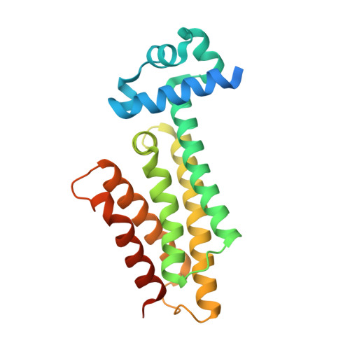
5MWO

Structure of Mycobacterium Tuberculosis Transcriptional Regulatory Repressor Protein (EthR) in complex with fragment 7E8.


Experimental Data Snapshot

  • Method: X-RAY DIFFRACTION
  • Resolution: 1.96 Å
  • R-Value Free: 0.259 
  • R-Value Work: 0.198 
  • R-Value Observed: 0.201 

Starting Model: experimental
View more details

wwPDB Validation   3D Report Full Report


Ligand Structure Quality Assessment 


This is version 1.3 of the entry. See complete history


Literature

Fragment Screening against the EthR-DNA Interaction by Native Mass Spectrometry.

Chan, D.S.Mendes, V.Thomas, S.E.McConnell, B.N.Matak-Vinkovic, D.Coyne, A.G.Blundell, T.L.Abell, C.

(2017) Angew Chem Int Ed Engl 56: 7488-7491

  • DOI: https://doi.org/10.1002/anie.201702888
  • Primary Citation of Related Structures:  
    5MWO, 5MXK

  • PubMed Abstract: 

    Native nanoelectrospray ionization mass spectrometry is an underutilized technique for fragment screening. In this study, the first demonstration is provided of the use of native mass spectrometry for screening fragments against a protein-DNA interaction. EthR is a transcriptional repressor of EthA expression in Mycobacterium tuberculosis (Mtb) that reduces the efficacy of ethionamide, a second-line antitubercular drug used to combat multidrug-resistant Mtb strains. A small-scale fragment screening campaign was conducted against the EthR-DNA interaction using native mass spectrometry, and the results were compared with those from differential scanning fluorimetry, a commonly used primary screening technique. Hits were validated by surface plasmon resonance and X-ray crystallography. The screening campaign identified two new fragments that disrupt the EthR-DNA interaction in vitro (IC 50 =460-610 μm) and bind to the hydrophobic channel of the EthR dimer.


  • Organizational Affiliation

    Department of Chemistry, University of Cambridge, Lensfield Road, Cambridge, CB2 1EW, UK.


Macromolecules
Find similar proteins by:  (by identity cutoff)  |  3D Structure
Entity ID: 1
MoleculeChains Sequence LengthOrganismDetailsImage
HTH-type transcriptional regulator EthR228Mycobacterium tuberculosis H37RvMutation(s): 0 
Gene Names: ethRetaRRv3855
UniProt
Find proteins for P9WMC1 (Mycobacterium tuberculosis (strain ATCC 25618 / H37Rv))
Explore P9WMC1 
Go to UniProtKB:  P9WMC1
Entity Groups  
Sequence Clusters30% Identity50% Identity70% Identity90% Identity95% Identity100% Identity
UniProt GroupP9WMC1
Sequence Annotations
Expand
  • Reference Sequence
Experimental Data & Validation

Experimental Data

  • Method: X-RAY DIFFRACTION
  • Resolution: 1.96 Å
  • R-Value Free: 0.259 
  • R-Value Work: 0.198 
  • R-Value Observed: 0.201 
  • Space Group: P 41 21 2
Unit Cell:
Length ( Å )Angle ( ˚ )
a = 121.927α = 90
b = 121.927β = 90
c = 33.728γ = 90
Software Package:
Software NamePurpose
PHENIXrefinement
XDSdata reduction
Aimlessdata scaling
PHASERphasing

Structure Validation

View Full Validation Report


Ligand Structure Quality Assessment 


Entry History & Funding Information

Deposition Data


Funding OrganizationLocationGrant Number
Bill & Melinda Gates FoundationUnited States--

Revision History  (Full details and data files)

  • Version 1.0: 2017-05-31
    Type: Initial release
  • Version 1.1: 2017-06-21
    Changes: Database references
  • Version 1.2: 2017-09-13
    Changes: Author supporting evidence
  • Version 1.3: 2024-01-17
    Changes: Data collection, Database references, Refinement description