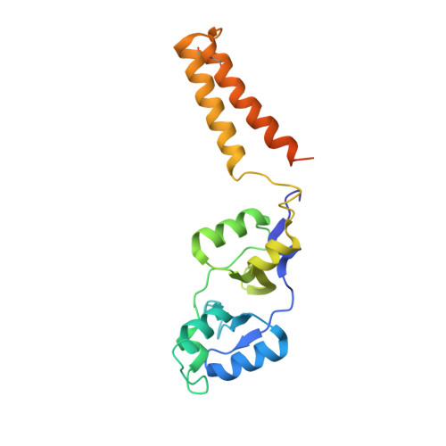
5NPL | pdb_00005npl

Crystal structure of hexameric CBS-CP12 protein from bloom-forming cyanobacteria, Yb-derivative at 2.8 A resolution


Experimental Data Snapshot

  • Method: X-RAY DIFFRACTION
  • Resolution: 2.79 Å
  • R-Value Free: 
    0.219 (Depositor), 0.220 (DCC) 
  • R-Value Work: 
    0.186 (Depositor), 0.200 (DCC) 
  • R-Value Observed: 
    0.188 (Depositor) 

wwPDB Validation   3D Report Full Report


Ligand Structure Quality Assessment 

Created with Raphaël 2.3.0Worse 01 BetterLigand structure goodness of fit to experimental dataBest fitted DO3Click on this verticalbar to view details

This is version 1.3 of the entry. See complete history


Literature

Structural and functional insights into the unique CBS-CP12 fusion protein family in cyanobacteria.

Hackenberg, C.Hakanpaa, J.Cai, F.Antonyuk, S.Eigner, C.Meissner, S.Laitaoja, M.Janis, J.Kerfeld, C.A.Dittmann, E.Lamzin, V.S.

(2018) Proc Natl Acad Sci U S A 115: 7141-7146

  • DOI: https://doi.org/10.1073/pnas.1806668115
  • Primary Citation of Related Structures:  
    5NMU, 5NPL, 5NVD

  • PubMed Abstract: 

    Cyanobacteria are important photosynthetic organisms inhabiting a range of dynamic environments. This phylum is distinctive among photosynthetic organisms in containing genes encoding uncharacterized cystathionine β-synthase (CBS)-chloroplast protein (CP12) fusion proteins. These consist of two domains, each recognized as stand-alone photosynthetic regulators with different functions described in cyanobacteria (CP12) and plants (CP12 and CBSX). Here we show that CBS-CP12 fusion proteins are encoded in distinct gene neighborhoods, several unrelated to photosynthesis. Most frequently, CBS-CP12 genes are in a gene cluster with thioredoxin A (TrxA), which is prevalent in bloom-forming, marine symbiotic, and benthic mat cyanobacteria. Focusing on a CBS-CP12 from Microcystis aeruginosa PCC 7806 encoded in a gene cluster with TrxA, we reveal that the domain fusion led to the formation of a hexameric protein. We show that the CP12 domain is essential for hexamerization and contains an ordered, previously structurally uncharacterized N-terminal region. We provide evidence that CBS-CP12, while combining properties of both regulatory domains, behaves different from CP12 and plant CBSX. It does not form a ternary complex with phosphoribulokinase (PRK) and glyceraldehyde-3-phosphate dehydrogenase. Instead, CBS-CP12 decreases the activity of PRK in an AMP-dependent manner. We propose that the novel domain architecture and oligomeric state of CBS-CP12 expand its regulatory function beyond those of CP12 in cyanobacteria.


  • Organizational Affiliation

    European Molecular Biology Laboratory (EMBL), Deutsches Elektronen-Synchrotron (DESY), 22607 Hamburg, Germany; hackenberg@embl-hamburg.de victor@embl-hamburg.de.


Macromolecules
Find similar proteins by:  (by identity cutoff)  |  3D Structure
Entity ID: 1
MoleculeChains Sequence LengthOrganismDetailsImage
Similar to tr|Q8YYT1|Q8YYT1
A, B, C
208Microcystis aeruginosa PCC 7806Mutation(s): 0 
Gene Names: IPF_2164
UniProt
Find proteins for A8YJ50 (Microcystis aeruginosa (strain PCC 7806))
Explore A8YJ50 
Go to UniProtKB:  A8YJ50
Entity Groups  
Sequence Clusters30% Identity50% Identity70% Identity90% Identity95% Identity100% Identity
UniProt GroupA8YJ50
Sequence Annotations
Expand
  • Reference Sequence
Small Molecules
Ligands 2 Unique
IDChains Name / Formula / InChI Key2D Diagram3D Interactions
DO3
Query on DO3

Download Ideal Coordinates CCD File 
E [auth A],
G [auth A]
10-((2R)-2-HYDROXYPROPYL)-1,4,7,10-TETRAAZACYCLODODECANE 1,4,7-TRIACETIC ACID
C17 H32 N4 O7
IQUHNCOJRJBMSU-CQSZACIVSA-N
YB
Query on YB

Download Ideal Coordinates CCD File 
D [auth A]
F [auth A]
H [auth A]
I [auth A]
J [auth A]
YTTERBIUM (III) ION
Yb
AWSFICBXMUKWSK-UHFFFAOYSA-N
Experimental Data & Validation

Experimental Data

  • Method: X-RAY DIFFRACTION
  • Resolution: 2.79 Å
  • R-Value Free:  0.219 (Depositor), 0.220 (DCC) 
  • R-Value Work:  0.186 (Depositor), 0.200 (DCC) 
  • R-Value Observed: 0.188 (Depositor) 
Space Group: C 2 2 21
Unit Cell:
Length ( Å )Angle ( ˚ )
a = 79.89α = 90
b = 120.734β = 90
c = 117.548γ = 90
Software Package:
Software NamePurpose
REFMACrefinement
XDSdata reduction
XDSdata scaling
CRANK2phasing

Structure Validation

View Full Validation Report


Ligand Structure Quality Assessment 

Created with Raphaël 2.3.0Worse 01 BetterLigand structure goodness of fit to experimental dataBest fitted DO3Click on this verticalbar to view details

Entry History 

Deposition Data

Revision History  (Full details and data files)

  • Version 1.0: 2018-05-30
    Type: Initial release
  • Version 1.1: 2018-06-20
    Changes: Data collection, Database references
  • Version 1.2: 2018-07-11
    Changes: Data collection, Database references
  • Version 1.3: 2024-11-06
    Changes: Data collection, Database references, Refinement description, Structure summary